

Volume 27, Issue 2 (7-2026)
Arch Rehabil 2026, 27(2): 326-341 |
Back to browse issues page



Ethics code: IR.MUBABOL.HRI.REC.1403.137


Download citation:
BibTeX | RIS | EndNote | Medlars | ProCite | Reference Manager | RefWorks
Send citation to:


BibTeX | RIS | EndNote | Medlars | ProCite | Reference Manager | RefWorks
Send citation to:
Elahi H, Talebi G A, Taghipour M, Nikbakht H A, Ebrahimi H. Comparison of Lumbar Lordosis and Pelvic Tilt Angles Between People With Chronic Non-specific Low Back Pain and Healthy Controls During Knee Flexion and Hip Extension. Arch Rehabil 2026; 27 (2) :326-341
URL: http://rehabilitationj.uswr.ac.ir/article-1-3672-en.html
URL: http://rehabilitationj.uswr.ac.ir/article-1-3672-en.html
Hossein Elahi1  , Ghadam Ali Talebi *2 , Mohammad Taghipour3 , Hossein Ali Nikbakht4 , Hadi Ebrahimi5
, Ghadam Ali Talebi *2 , Mohammad Taghipour3 , Hossein Ali Nikbakht4 , Hadi Ebrahimi5
1- Department of Physiotherapy Student Research Committee, Babol University of Medical Science, Babol, Iran.
2- Mobility Impairment Research Center, Health Research Institute, Babol University of Medical Sciences, Babol, Iran. ,Talebiali2@yahoo.co.in
3- Mobility Impairment Research Center, Health Research Institute, Babol University of Medical Sciences, Babol, Iran.
4- Social Determinants of Health Research Center, Health Research Institute, Babol University of Medical Sciences, Babol, Iran.
5- Clinical Research Development Unit of Shahid Beheshti Hospital, Babol University of Medical Sciences, Babol, Iran.
2- Mobility Impairment Research Center, Health Research Institute, Babol University of Medical Sciences, Babol, Iran. ,
3- Mobility Impairment Research Center, Health Research Institute, Babol University of Medical Sciences, Babol, Iran.
4- Social Determinants of Health Research Center, Health Research Institute, Babol University of Medical Sciences, Babol, Iran.
5- Clinical Research Development Unit of Shahid Beheshti Hospital, Babol University of Medical Sciences, Babol, Iran.
Full-Text [PDF 1750 kb]
(186 Downloads)
| Abstract (HTML) (2396 Views)
Full-Text: (204 Views)
Introduction
Low back pain (LBP) is a common musculoskeletal issue that causes individuals to visit physiotherapy centers [1, 2]. The prevalence of LBP ranges from 30% to 80% and increases with aging [3]. LBP is categorized into two types: specific (caused by specific pathophysiological mechanisms) and non-specific (with no definitive cause). Non-specific LBP constitutes approximately 80-90% of all LBP cases. Moreover, based on duration, LBP can be acute (<6 weeks), subacute (6-12 weeks), or chronic (>12 weeks) [4]. Both lumbar lordosis and pelvic tilt exhibit dynamic behavior, adapting to various influences such as spinal pathology or dysfunction in adjacent joints. In LBP patients, these parameters may respond differently to lower limb movements, including hip extension and knee flexion, than in healthy individuals. This different response aligns with Sahrmann’s concept of relative flexibility, which posits that incorrect lumbopelvic alignment or compensatory movement patterns contribute to the occurrence of pain syndrome [5]. In biomechanical evaluation of LBP, lumbar lordosis angle (LLA) is usually considered as a primary measure. The active knee flexion test and the active hip extension test in the prone position are two standard clinical measures for evaluating lumbar movement impairment [5].
The existing literature presents mixed results regarding comparisons of LLA and pelvic tilt between LBP patients and healthy individuals, as well as the effects of active hip extension and knee flexion tests on these variables. Singh et al. [6], Harrison et al. [7], and Ifthekar et al. [8] reported no significant differences in LLA between patients with LBP and asymptomatic controls. Similarly, Arab et al. found that differences in LLA adjustments following a prone knee flexion test were not statistically significant between healthy individuals and LBP subjects [9]. In another study, however, they observed significant differences in LLA changes following the prone hip extension test between the two groups [10]. Evidence from systematic reviews shows conflicting findings regarding the association between LLA and the development of LBP. In a meta-analysis of 13 studies comprising 796 patients with LBP and 927 healthy individuals, conducted by Chun et al., patients with LBP exhibited a lower LLA than healthy individuals. Subgroup analyses indicated that subjects with lumbar disc herniation or degenerative spinal changes demonstrated significantly lower LLA than controls. However, the included studies exhibited considerable heterogeneity in terms of methodology and sample characteristics [11]. Lim et al. showed a small but statistically significant difference in pelvic tilt angle (PTA) between healthy controls and LBP patients [12]. Conversely, Yasukouchi et al. found no significant differences in PTA changes in various standing and sitting positions when comparing healthy subjects to those with LBP [13].
Several key considerations support the rationale for this study. First, the reports linking PTA, LLA, and LBP symptoms are contradictory. Both increased and decreased LLA have been reported in patients with specific LBP [11]. However, in patients with chronic non-specific LBP (CNSLBP), a mixed condition that combines different syndromes (restrictive dysfunction, functional instability, and flexion or extension movement deficits), the link remains unclear and warrants further research [2]. It is also unclear whether the duration or severity of CNSLBP influences LLA, underscoring the need for additional research into this potential association [3]. From a theoretical perspective, individuals with LBP may exhibit altered LLA during active knee flexion or hip extension due to stiffness or compensatory lumbopelvic movements. However, the simultaneous effects of these movements on LLA and PTA have not yet been evaluated in either the prone or standing position. Therefore, this study aimed to evaluate and compare LLA changes during prone hip extension and knee flexion tests between CNSLBP patients and asymptomatic controls. The study also compared PTA changes in the standing position between these two groups. The analysis examined how pain duration and symptom severity correlated with PTA and LLA in patients. The results can be used in the orthopedic evaluation and rehabilitation program of patients with CNSLBP.
Materials and Methods
Study design and participants
This is a case-control study conducted at Shahid Beheshti Hospital, affiliated with Babol University of Medical Sciences, Babol, Iran. The case group comprised individuals with CNSLBP who attended orthopedic or physiotherapy clinics of Shahid Beheshti Hospital between October 2023 and March 2024. The control group consisted of healthy volunteers without LBP, verified through self-report and clinical examination. Participants were selected via convenience sampling from individuals who met the inclusion and exclusion criteria. To address the confounding factors, the two groups were matched for age and body weight. Inclusion criteria were: age 20-45 years, confirmed diagnosis of CNSLBP by an orthopedic surgeon for the case group [10, 14], and no history of LBP for the control group. Exclusion criteria were: history of breathing difficulties in the prone position, history of dislocation or fracture, acute LBP with pain intensity ≥8 on the visual analog scale (VAS) score. confirmed disc herniation or spinal canal stenosis, previous spinal surgery, history of anterior cruciate ligament injury, history of anterior knee pain, leg length discrepancy exceeding 1 cm, inability to perform active hip extension or knee flexion, lower limb injury within the past three months, presence of positive neurological signs, and inability to maintain cooperation or continue participation in the study. G*Power software (version 3) was used to calculate the sample size considering an effect size of 0.68 based on prior research [10], a 95% confidence level, and a test power of 95%. The sample size was obtained at 56 per group (a total of 112 individuals). Considering a potential 10% dropout, the final total size was 124 (62 per group).
Procedure
Orthopedic specialists examined the patients and healthy volunteers. Included participants provided written informed consent before the study, and the Ethics Committee of Babol University of Medical Sciences approved the research proposal. Initial steps included gathering demographic information and assessing pain duration and symptom intensity (using the VAS) in clinical cases.
The LLA was measured in both groups using a flexible ruler, first in a prone position and then immediately after active hip extension (up to 10°) and active knee flexion. The purpose of these two maneuvers was to evaluate the influence of compensatory relative flexibility on lumbar spine behavior. Accordingly, lumbopelvic responses during these tasks were compared between case and control groups. The measurement began by identifying the spinous process of the twelfth thoracic vertebra (T12) as the starting point and the spinous process of the second sacral vertebra (S2) as the endpoint of the lumbar curve. These anatomical landmarks were marked on the participant’s skin, and the flexible ruler was then molded along the curvature of the lumbar spine. Contact points on the ruler corresponding to the midline marks were indicated with a marker pen. The LLA (θ) was calculated using the following Equation 1: [15, 16], Where L is the length between T12 and S2, and H is the maximum height of the lumbar curve [9, 10].
1. θ=4× arctan(2H/L)
An inclinometer (made in Belgium) was used to measure the standing PTA. Subjects stood straight during device placement between the anterior superior iliac spine and the posterior superior iliac spine. The instrument screen showed the PTA [15]. The prone LLA and standing PTA were measured three times in three conditions: relaxed position, immediately after active knee flexion, and immediately after active hip extension. The average of three measurements per condition was used in the data analysis.
Data analysis
The normal distribution of the data was confirmed using the Kolmogorov-Smirnov test. Independent t-test compared the mean PTA and LLA between the two groups. One-way analysis of variance (ANOVA) evaluated within-group differences across the three conditions. Pearson’s correlation test assessed whether pain severity or symptom duration were associated with PTA and LLA in the case group. Mean difference and 95% confidence interval (CI) were used to show the effect sizes. Calculations were performed in SPSS software, version 23, with a statistical significance level of 0.05. The intraclass correlation coefficient (ICC) was used to assess the reliability of measurements obtained from 10 participants.
Results
Participants were 62 CNSLBP patients (mean age: 36.55 years, mean weight: 75.04 kg) and 62 asymptomatic peers (mean age: 35.86 years, mean weight: 75.19 kg). Chi-square test results showed no significant difference in sex between the two groups (P=0.999). Other demographic baselines also showed no statistically significant differences between the two groups (P>0.05). The ICC for all PTA and LLA exceeded 0.9, confirming strong measurement reliability.
According to the results in Table 1, in the case group, the highest mean LLA was obtained during hip extension (38.03±12.75 degrees), while in the control group, the highest value was during knee flexion (36.38±11.24 degrees).
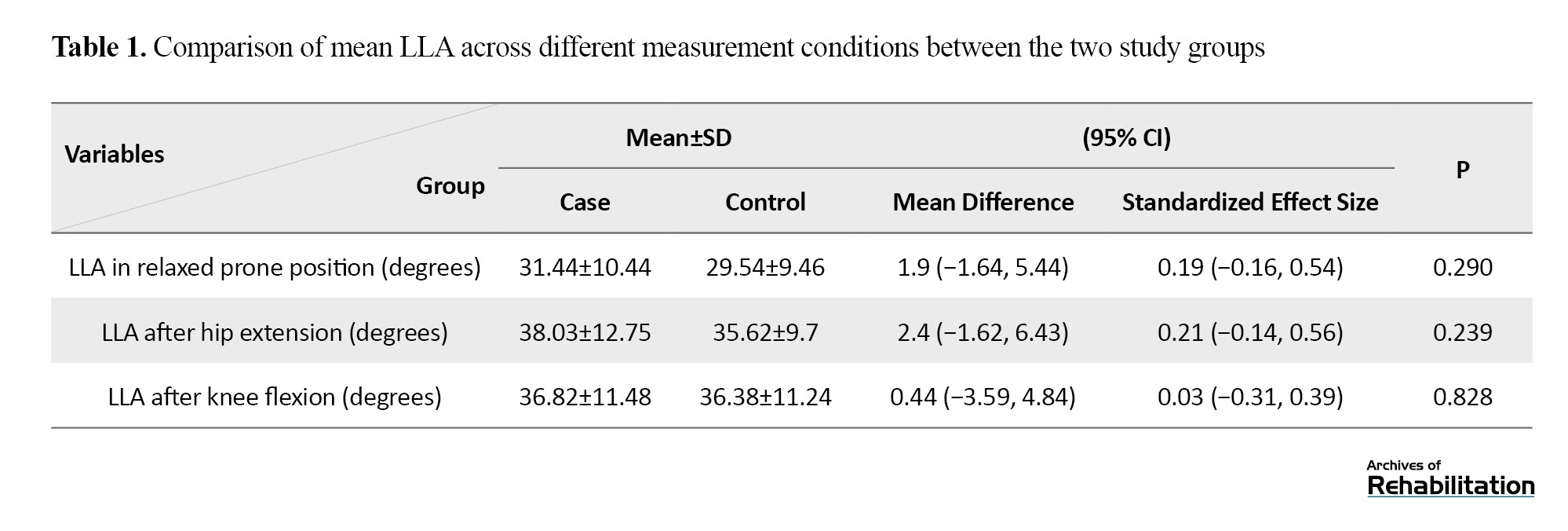
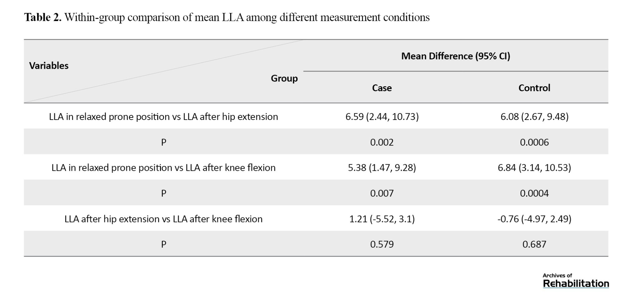
Mean LLA was not significantly different between the two groups across different measurement conditions (P>0.05). Standardized effect sizes indicated minor differences between groups. Within-group analysis revealed that LLA values after hip extension and knee flexion were significantly greater than in the relaxed prone condition in both groups (P<0.001) (Table 2).
In both groups, the highest mean PTA was observed after hip extension (11.03 ± 5.88 degrees in the case group and 12.74±5.48 degrees in the control group (Table 3).
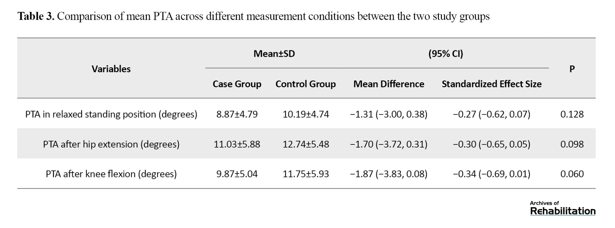
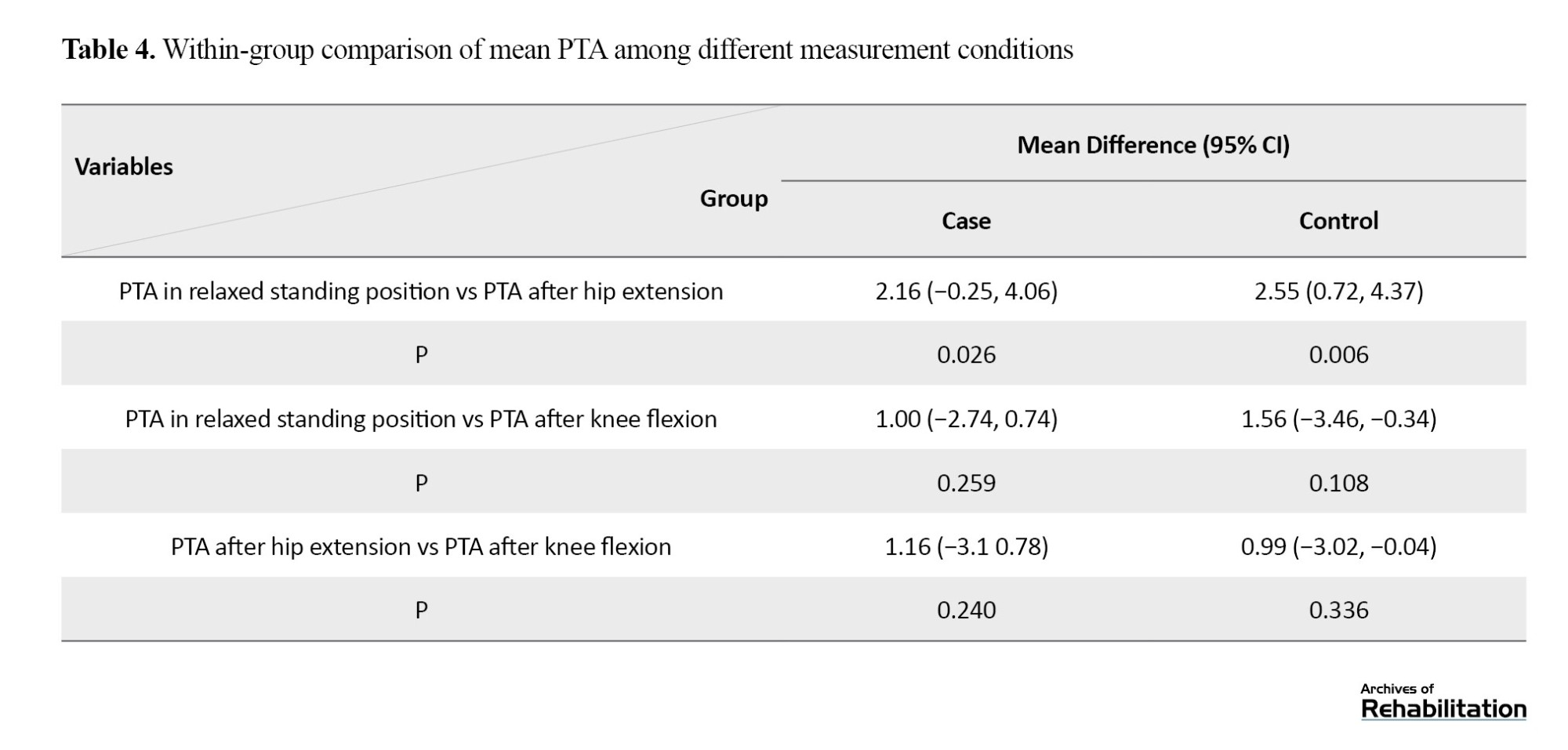
Analysis of PTA in the standing position showed no significant differences between groups across different conditions (P>0.05). Effect sizes again suggested minimal differences. Within-group comparisons indicated that PTA after hip extension was significantly higher compared to PTA in the relaxed standing position in both groups (P<0.05) (Table 4).
Symptom duration and pain intensity demonstrated no significant correlations with PTA or LLA across most conditions. A weak positive and significant correlation was found only between pain intensity and standing PTA after hip extension (P=0.022).
Discussion
In the present study, LLA was significantly higher after active hip extension and knee flexion than in the relaxed prone position in both case and control groups. However, the results showed no significant differences among the three measurement conditions.
Arab et al. also reported that changes in LLA during the prone knee flexion test were not statistically significant between LBP patients and healthy peers. However, the patient group exhibited more changes [10]. Conversely, Arab et al. in another study found a significant difference between the two groups in LLA changes when comparing the relaxed prone and post hip extension conditions [9]. Singh et al. [6], Harrison et al. [7], and Ifthekar et al. identified no significant differences in LLA between LBP patients and healthy controls [8]. These contradictory findings suggest that LLA alone may not be a reliable discriminator between individuals with and without CNSLBP during movement tests. It is likely that other factors such as neuromuscular control, movement coordination, and individual biomechanical variations play more significant roles in the pathophysiology of CNSLBP. Future studies incorporating dynamic assessments and comprehensive biomechanical analyses may provide deeper insights.
This study showed that, in both groups, mean PTA after hip extension was significantly greater than that in the relaxed standing position. Direct group comparisons between healthy individuals and patients revealed no significant differences in PTA among any measurement conditions. Lim et al. reported a small but statistically significant difference in PTA between patients with LBP and healthy controls [12]. Day et al. also reported that PTA variations in LBP patients could lead to significant changes in the overall pelvic and lumbar curves [17]. In contrast, Yasukouchi et al. documented identical pelvic tilt adaptations in sitting and standing positions between patients with LBP and healthy controls [13]. Adamik et al. reported reduced anterior PTA in patients with LBP [18].
This study evaluated whether LBP intensity or symptom duration was directly related to PTA and LLA values. Standing PTA after hip extension demonstrated a weak, statistically significant positive correlation with overall pain intensity. The weak correlation may be related to the generally greater PTA observed in the standing hip extension compared to other measurement conditions. Shortz et al. [19] and Singh et al. [6] identified no significant correlation between LLA and chronic LBP severity. Bozorgmehr et al. reported a significant positive correlation between pain severity and PTA alterations [20]. Elabd and Elabd identified a significant correlation between pain severity and LLA, and Leo et al. found significant positive correlations between pain intensity and alterations in both LLA and PTA in patients with CNSLBP [21, 22].
The present findings show that LLA and PTA across three different measurement conditions do not differ significantly between healthy controls and CNSLBP patients. Therefore, the presence of LBP seems insufficient to determine the changes in LLA and PTA in patients. In a systematic review by Chun et al., a decrease in LLA was reported in subjects with LBP compared with healthy controls. The authors suggested that decreased LLA, often associated with sustained lumbar flexion, may contribute to posterior disc protrusion or herniation. In the present study, participants had CNSLBP. These people may suffer from a broad range of musculoskeletal pain syndromes, including restrictive movement disorders, extension and flexion impairment, or functional instability. The presence of various movement disorders in people with CNSLBP likely results in different effects on postural alignment of the lumbar spine and pelvis. Patients in our study exhibited both increased and decreased LLA. Consequently, the absence of significant differences in mean LLA and PTA between CNSLBP patients and asymptomatic controls may be due to the heterogeneity of movement disorders among CNSLBP patients [11].
Conclusion
To better elucidate the relationship between lumbopelvic alignment disorders and LBP, future research should employ larger sample sizes and focus on distinct subgroups of people with CNSLBP, particularly those with defined movement impairment syndromes, such as extension or flexion dysfunctions. Furthermore, studies examining changes in lumbar curvature and pelvic tilt in this population should control for a range of potentially influential variables, including age, symptom duration and severity, specific characteristics of movement impairment, and musculoskeletal and neuromuscular elements (e.g. muscle length, tightness, stiffness, strength, endurance), as well as patterns of relative flexibility and compensatory movement strategies. Based on the findings, although LLA and PTA are frequently assessed as primary postural indicators during orthopedic evaluations of patients with LPB, clinicians should not overemphasize the role of these parameters without considering other biomechanical and neuromuscular factors.
There were some limitations in this study. A small sample size may limit the generalizability of the findings to broader clinical populations. The non-assessment of other potentially influential factors, such as muscle strength, core endurance, muscle length, tightness, stiffness, and neuromuscular compensatory patterns, limits a comprehensive understanding of the underlying mechanisms. The lack of sophisticated radiological tools such as MRI may affect the accuracy of LLA and PTA measurements.
Ethical Considerations
Compliance with ethical guidelines
This study was approved by the Research Ethics Committee of Babol University of Medical Sciences, Babol, Iran (Code: IR.MUBABOL.HRI.REC.1403.137). Participants provided written informed consent prior to data collection.
Funding
This research did not receive any grant from funding agencies in the public, commercial, or non-profit sectors.
Authors' contributions
Conceptualization: Hossein Elahi and Ghadam Ali Talebi; Methodology and data analysis: Hossein-Ali Nikbakht and Ghadam Ali Talebi; Investigations: Mohammad Taghipour and Hadi Ebrahimi; Writing the initial draft and supervision: Ghadam Ali Talebi; Review and editing: All authors.
Conflict of interest
The authors declared no conflict of interest.
AI tools disclosure statement
The authors acknowledge that no AI tools were used in the preparation of this article.
Acknowledgments
The authors sincerely thank the staff and personnel of the physiotherapy and orthopedic clinics at Shahid Beheshti Hospital in Babol, as well as all participants, for their time and cooperation.
Low back pain (LBP) is a common musculoskeletal issue that causes individuals to visit physiotherapy centers [1, 2]. The prevalence of LBP ranges from 30% to 80% and increases with aging [3]. LBP is categorized into two types: specific (caused by specific pathophysiological mechanisms) and non-specific (with no definitive cause). Non-specific LBP constitutes approximately 80-90% of all LBP cases. Moreover, based on duration, LBP can be acute (<6 weeks), subacute (6-12 weeks), or chronic (>12 weeks) [4]. Both lumbar lordosis and pelvic tilt exhibit dynamic behavior, adapting to various influences such as spinal pathology or dysfunction in adjacent joints. In LBP patients, these parameters may respond differently to lower limb movements, including hip extension and knee flexion, than in healthy individuals. This different response aligns with Sahrmann’s concept of relative flexibility, which posits that incorrect lumbopelvic alignment or compensatory movement patterns contribute to the occurrence of pain syndrome [5]. In biomechanical evaluation of LBP, lumbar lordosis angle (LLA) is usually considered as a primary measure. The active knee flexion test and the active hip extension test in the prone position are two standard clinical measures for evaluating lumbar movement impairment [5].
The existing literature presents mixed results regarding comparisons of LLA and pelvic tilt between LBP patients and healthy individuals, as well as the effects of active hip extension and knee flexion tests on these variables. Singh et al. [6], Harrison et al. [7], and Ifthekar et al. [8] reported no significant differences in LLA between patients with LBP and asymptomatic controls. Similarly, Arab et al. found that differences in LLA adjustments following a prone knee flexion test were not statistically significant between healthy individuals and LBP subjects [9]. In another study, however, they observed significant differences in LLA changes following the prone hip extension test between the two groups [10]. Evidence from systematic reviews shows conflicting findings regarding the association between LLA and the development of LBP. In a meta-analysis of 13 studies comprising 796 patients with LBP and 927 healthy individuals, conducted by Chun et al., patients with LBP exhibited a lower LLA than healthy individuals. Subgroup analyses indicated that subjects with lumbar disc herniation or degenerative spinal changes demonstrated significantly lower LLA than controls. However, the included studies exhibited considerable heterogeneity in terms of methodology and sample characteristics [11]. Lim et al. showed a small but statistically significant difference in pelvic tilt angle (PTA) between healthy controls and LBP patients [12]. Conversely, Yasukouchi et al. found no significant differences in PTA changes in various standing and sitting positions when comparing healthy subjects to those with LBP [13].
Several key considerations support the rationale for this study. First, the reports linking PTA, LLA, and LBP symptoms are contradictory. Both increased and decreased LLA have been reported in patients with specific LBP [11]. However, in patients with chronic non-specific LBP (CNSLBP), a mixed condition that combines different syndromes (restrictive dysfunction, functional instability, and flexion or extension movement deficits), the link remains unclear and warrants further research [2]. It is also unclear whether the duration or severity of CNSLBP influences LLA, underscoring the need for additional research into this potential association [3]. From a theoretical perspective, individuals with LBP may exhibit altered LLA during active knee flexion or hip extension due to stiffness or compensatory lumbopelvic movements. However, the simultaneous effects of these movements on LLA and PTA have not yet been evaluated in either the prone or standing position. Therefore, this study aimed to evaluate and compare LLA changes during prone hip extension and knee flexion tests between CNSLBP patients and asymptomatic controls. The study also compared PTA changes in the standing position between these two groups. The analysis examined how pain duration and symptom severity correlated with PTA and LLA in patients. The results can be used in the orthopedic evaluation and rehabilitation program of patients with CNSLBP.
Materials and Methods
Study design and participants
This is a case-control study conducted at Shahid Beheshti Hospital, affiliated with Babol University of Medical Sciences, Babol, Iran. The case group comprised individuals with CNSLBP who attended orthopedic or physiotherapy clinics of Shahid Beheshti Hospital between October 2023 and March 2024. The control group consisted of healthy volunteers without LBP, verified through self-report and clinical examination. Participants were selected via convenience sampling from individuals who met the inclusion and exclusion criteria. To address the confounding factors, the two groups were matched for age and body weight. Inclusion criteria were: age 20-45 years, confirmed diagnosis of CNSLBP by an orthopedic surgeon for the case group [10, 14], and no history of LBP for the control group. Exclusion criteria were: history of breathing difficulties in the prone position, history of dislocation or fracture, acute LBP with pain intensity ≥8 on the visual analog scale (VAS) score. confirmed disc herniation or spinal canal stenosis, previous spinal surgery, history of anterior cruciate ligament injury, history of anterior knee pain, leg length discrepancy exceeding 1 cm, inability to perform active hip extension or knee flexion, lower limb injury within the past three months, presence of positive neurological signs, and inability to maintain cooperation or continue participation in the study. G*Power software (version 3) was used to calculate the sample size considering an effect size of 0.68 based on prior research [10], a 95% confidence level, and a test power of 95%. The sample size was obtained at 56 per group (a total of 112 individuals). Considering a potential 10% dropout, the final total size was 124 (62 per group).
Procedure
Orthopedic specialists examined the patients and healthy volunteers. Included participants provided written informed consent before the study, and the Ethics Committee of Babol University of Medical Sciences approved the research proposal. Initial steps included gathering demographic information and assessing pain duration and symptom intensity (using the VAS) in clinical cases.
The LLA was measured in both groups using a flexible ruler, first in a prone position and then immediately after active hip extension (up to 10°) and active knee flexion. The purpose of these two maneuvers was to evaluate the influence of compensatory relative flexibility on lumbar spine behavior. Accordingly, lumbopelvic responses during these tasks were compared between case and control groups. The measurement began by identifying the spinous process of the twelfth thoracic vertebra (T12) as the starting point and the spinous process of the second sacral vertebra (S2) as the endpoint of the lumbar curve. These anatomical landmarks were marked on the participant’s skin, and the flexible ruler was then molded along the curvature of the lumbar spine. Contact points on the ruler corresponding to the midline marks were indicated with a marker pen. The LLA (θ) was calculated using the following Equation 1: [15, 16], Where L is the length between T12 and S2, and H is the maximum height of the lumbar curve [9, 10].
1. θ=4× arctan(2H/L)
An inclinometer (made in Belgium) was used to measure the standing PTA. Subjects stood straight during device placement between the anterior superior iliac spine and the posterior superior iliac spine. The instrument screen showed the PTA [15]. The prone LLA and standing PTA were measured three times in three conditions: relaxed position, immediately after active knee flexion, and immediately after active hip extension. The average of three measurements per condition was used in the data analysis.
Data analysis
The normal distribution of the data was confirmed using the Kolmogorov-Smirnov test. Independent t-test compared the mean PTA and LLA between the two groups. One-way analysis of variance (ANOVA) evaluated within-group differences across the three conditions. Pearson’s correlation test assessed whether pain severity or symptom duration were associated with PTA and LLA in the case group. Mean difference and 95% confidence interval (CI) were used to show the effect sizes. Calculations were performed in SPSS software, version 23, with a statistical significance level of 0.05. The intraclass correlation coefficient (ICC) was used to assess the reliability of measurements obtained from 10 participants.
Results
Participants were 62 CNSLBP patients (mean age: 36.55 years, mean weight: 75.04 kg) and 62 asymptomatic peers (mean age: 35.86 years, mean weight: 75.19 kg). Chi-square test results showed no significant difference in sex between the two groups (P=0.999). Other demographic baselines also showed no statistically significant differences between the two groups (P>0.05). The ICC for all PTA and LLA exceeded 0.9, confirming strong measurement reliability.
According to the results in Table 1, in the case group, the highest mean LLA was obtained during hip extension (38.03±12.75 degrees), while in the control group, the highest value was during knee flexion (36.38±11.24 degrees).
Mean LLA was not significantly different between the two groups across different measurement conditions (P>0.05). Standardized effect sizes indicated minor differences between groups. Within-group analysis revealed that LLA values after hip extension and knee flexion were significantly greater than in the relaxed prone condition in both groups (P<0.001) (Table 2).
In both groups, the highest mean PTA was observed after hip extension (11.03 ± 5.88 degrees in the case group and 12.74±5.48 degrees in the control group (Table 3).
Analysis of PTA in the standing position showed no significant differences between groups across different conditions (P>0.05). Effect sizes again suggested minimal differences. Within-group comparisons indicated that PTA after hip extension was significantly higher compared to PTA in the relaxed standing position in both groups (P<0.05) (Table 4).
Symptom duration and pain intensity demonstrated no significant correlations with PTA or LLA across most conditions. A weak positive and significant correlation was found only between pain intensity and standing PTA after hip extension (P=0.022).
Discussion
In the present study, LLA was significantly higher after active hip extension and knee flexion than in the relaxed prone position in both case and control groups. However, the results showed no significant differences among the three measurement conditions.
Arab et al. also reported that changes in LLA during the prone knee flexion test were not statistically significant between LBP patients and healthy peers. However, the patient group exhibited more changes [10]. Conversely, Arab et al. in another study found a significant difference between the two groups in LLA changes when comparing the relaxed prone and post hip extension conditions [9]. Singh et al. [6], Harrison et al. [7], and Ifthekar et al. identified no significant differences in LLA between LBP patients and healthy controls [8]. These contradictory findings suggest that LLA alone may not be a reliable discriminator between individuals with and without CNSLBP during movement tests. It is likely that other factors such as neuromuscular control, movement coordination, and individual biomechanical variations play more significant roles in the pathophysiology of CNSLBP. Future studies incorporating dynamic assessments and comprehensive biomechanical analyses may provide deeper insights.
This study showed that, in both groups, mean PTA after hip extension was significantly greater than that in the relaxed standing position. Direct group comparisons between healthy individuals and patients revealed no significant differences in PTA among any measurement conditions. Lim et al. reported a small but statistically significant difference in PTA between patients with LBP and healthy controls [12]. Day et al. also reported that PTA variations in LBP patients could lead to significant changes in the overall pelvic and lumbar curves [17]. In contrast, Yasukouchi et al. documented identical pelvic tilt adaptations in sitting and standing positions between patients with LBP and healthy controls [13]. Adamik et al. reported reduced anterior PTA in patients with LBP [18].
This study evaluated whether LBP intensity or symptom duration was directly related to PTA and LLA values. Standing PTA after hip extension demonstrated a weak, statistically significant positive correlation with overall pain intensity. The weak correlation may be related to the generally greater PTA observed in the standing hip extension compared to other measurement conditions. Shortz et al. [19] and Singh et al. [6] identified no significant correlation between LLA and chronic LBP severity. Bozorgmehr et al. reported a significant positive correlation between pain severity and PTA alterations [20]. Elabd and Elabd identified a significant correlation between pain severity and LLA, and Leo et al. found significant positive correlations between pain intensity and alterations in both LLA and PTA in patients with CNSLBP [21, 22].
The present findings show that LLA and PTA across three different measurement conditions do not differ significantly between healthy controls and CNSLBP patients. Therefore, the presence of LBP seems insufficient to determine the changes in LLA and PTA in patients. In a systematic review by Chun et al., a decrease in LLA was reported in subjects with LBP compared with healthy controls. The authors suggested that decreased LLA, often associated with sustained lumbar flexion, may contribute to posterior disc protrusion or herniation. In the present study, participants had CNSLBP. These people may suffer from a broad range of musculoskeletal pain syndromes, including restrictive movement disorders, extension and flexion impairment, or functional instability. The presence of various movement disorders in people with CNSLBP likely results in different effects on postural alignment of the lumbar spine and pelvis. Patients in our study exhibited both increased and decreased LLA. Consequently, the absence of significant differences in mean LLA and PTA between CNSLBP patients and asymptomatic controls may be due to the heterogeneity of movement disorders among CNSLBP patients [11].
Conclusion
To better elucidate the relationship between lumbopelvic alignment disorders and LBP, future research should employ larger sample sizes and focus on distinct subgroups of people with CNSLBP, particularly those with defined movement impairment syndromes, such as extension or flexion dysfunctions. Furthermore, studies examining changes in lumbar curvature and pelvic tilt in this population should control for a range of potentially influential variables, including age, symptom duration and severity, specific characteristics of movement impairment, and musculoskeletal and neuromuscular elements (e.g. muscle length, tightness, stiffness, strength, endurance), as well as patterns of relative flexibility and compensatory movement strategies. Based on the findings, although LLA and PTA are frequently assessed as primary postural indicators during orthopedic evaluations of patients with LPB, clinicians should not overemphasize the role of these parameters without considering other biomechanical and neuromuscular factors.
There were some limitations in this study. A small sample size may limit the generalizability of the findings to broader clinical populations. The non-assessment of other potentially influential factors, such as muscle strength, core endurance, muscle length, tightness, stiffness, and neuromuscular compensatory patterns, limits a comprehensive understanding of the underlying mechanisms. The lack of sophisticated radiological tools such as MRI may affect the accuracy of LLA and PTA measurements.
Ethical Considerations
Compliance with ethical guidelines
This study was approved by the Research Ethics Committee of Babol University of Medical Sciences, Babol, Iran (Code: IR.MUBABOL.HRI.REC.1403.137). Participants provided written informed consent prior to data collection.
Funding
This research did not receive any grant from funding agencies in the public, commercial, or non-profit sectors.
Authors' contributions
Conceptualization: Hossein Elahi and Ghadam Ali Talebi; Methodology and data analysis: Hossein-Ali Nikbakht and Ghadam Ali Talebi; Investigations: Mohammad Taghipour and Hadi Ebrahimi; Writing the initial draft and supervision: Ghadam Ali Talebi; Review and editing: All authors.
Conflict of interest
The authors declared no conflict of interest.
AI tools disclosure statement
The authors acknowledge that no AI tools were used in the preparation of this article.
Acknowledgments
The authors sincerely thank the staff and personnel of the physiotherapy and orthopedic clinics at Shahid Beheshti Hospital in Babol, as well as all participants, for their time and cooperation.
References
- Rechtine GR. Diagnosis and treatment of low back pain. Journal of Orthopaedic Trauma. 1992;6(3):395. [DOI:10.1097/00005131-199209000-00031]
- Cieza A, Causey K, Kamenov K, Hanson SW, Chatterji S, Vos T. Global estimates of the need for rehabilitation based on the Global Burden of Disease study 2019: a systematic analysis for the Global Burden of Disease Study 2019. The Lancet. 2020;396(10267):2006-17. [DOI:10.1016/S0140-6736(20)32340-0] [PMID] [PMCID]
- Shokri P, Zahmatyar M, Falah Tafti M, Fathy M, Rezaei Tolzali M, Ghaffari Jolfayi A, et al. Non-spinal low back pain: Global epidemiology, trends, and risk factors. Health Science Reports. 2023;6(9):e1533. [DOI:10.1002/hsr2.1533] [PMID] [PMCID]
- Chiarotto A, Koes BW. Nonspecific low back pain. New England Journal of Medicine. 2022;386(18):1732-40. [DOI:10.1056/NEJMcp2032396] [PMID]
- Sahrmann S. Diagnosis and treatment of movement impairment syndromes-e-book: Elsevier Health Sciences; 2001.
- Singh S, Singh VP, Jain S. Relationship of Lumbar Lordosis With Non-specific Acute Low Back Pain. Cureus. 2024;16(10):e72453. [DOI:10.7759/cureus.72453]
- Harrison DE, Haas JW, Moustafa IM, Betz JW, Oakley PA. Can the Mismatch of Measured Pelvic Morphology vs Lumbar Lordosis Predict Chronic Low Back Pain Patients? Journal of Clinical Medicine. 2024;13(8):2178. [DOI:10.3390/jcm13082178] [PMID] [PMCID]
- Ifthekar S, Yadav G, Ahuja K, Mittal S, Venkata SP, Kandwal P. Correlation of spinopelvic parameters with functional outcomes in surgically managed cases of lumbar spinal tuberculosis-A retrospective study. Journal of Clinical Orthopaedics and Trauma. 2022;26:101788. [DOI:10.1016/j.jcot.2022.101788] [PMID] [PMCID]
- Arab AM, Talimkhani A, Karimi N, Ehsani F. Change in lumbar lordosis during prone lying knee flexion test in subjects with and without low back pain. Chiropractic & Manual Therapies. 2015;23:1-5. [DOI:10.1186/s12998-015-0061-z] [PMID] [PMCID]
- Arab AM, Haghighat A, Amiri Z, Khosravi F. Lumbar lordosis in prone position and prone hip extension test: comparison between subjects with and without low back pain. Chiropr Man Therap. 2017;25:8. [DOI:10.1186/s12998-017-0139-x] [PMID] [PMCID]
- Chun SW, Lim CY, Kim K, Hwang J, Chung SG. The relationships between low back pain and lumbar lordosis: a systematic review and meta-analysis. Spine J. 2017;17(8):1180-91. [DOI:10.1016/j.spinee.2017.04.034] [PMID]
- Lim HS, Roh SY, Lee SM. The Relationship between Pelvic Tilt Angle and Disability Associated with Low Back Pain. Journal of Physical Therapy Science. 2013;25(1):65-8. [DOI:10.1589/jpts.25.65]
- Yasukouchi A, Isayama T. The relationships between lumbar curves, pelvic tilt and joint mobilities in different sitting postures in young adult males. Applied human science. 1995;14(1):15-21. [DOI:10.2114/ahs.14.15] [PMID]
- Choi W-J, Kim W-D, Park D-C, Shin D-C. Comparison of compensatory lumbar movement in participants with and without non-specific chronic low back pain: a cross-sectional study. Journal of Back and Musculoskeletal Rehabilitation. 2022;35(6):1365-72. [DOI:10.3233/BMR-210327] [PMID]
- Naseri N, Fakhari Z, Haji Maghsoudi M, Hosseini Ghahi F, Senobari M. The relationship between pelvic tilt and lumbar lordosis among healthy females of 20 to 65 years. Modern Rehabilitation. 2014;8(3):76-81.
- Hart DL, Rose SJ. Reliability of a noninvasive method for measuring the lumbar curve*. J Orthop Sports Phys Ther. 1986;8(4):180-4. [DOI:10.2519/jospt.1986.8.4.180] [PMID]
- Day JW, Smidt GL, Lehmann T. Effect of pelvic tilt on standing posture. Physical Therapy. 1984;64(4):510-6. [DOI:10.1093/ptj/64.4.510] [PMID]
- Adamik A, Mikołajczyk E. Functional Patterns and Postural Balance Among Young Women with Chronic Non-Specific Low Back Pain and in Healthy Individuals. Medical Rehabilitation. 2024;28(2):23-30. [DOI:10.5604/01.3001.0054.6088]
- Shortz SK, Haas M. Relationship between radiographic lumbosacral spine mensuration and chronic low back pain intensity: a cross-sectional study. Journal of chiropractic medicine. 2018;17(1):1-6. [DOI:10.1016/j.jcm.2017.10.005] [PMID] [PMCID]
- Bozorgmehr A, Takamjani IE, Akbari M, Salehi R, Mohsenifar H, Rasouli O. Effect of posterior pelvic tilt taping on abdominal muscle thickness and lumbar lordosis in individuals with chronic low back pain and hyperlordosis: A single-group, repeated-measures trial. Journal of chiropractic medicine. 2020;19(4):213-21. [DOI:10.1016/j.jcm.2020.07.001] [PMID] [PMCID]
- Elabd AM, Elabd OM. Relationships between forward head posture and lumbopelvic sagittal alignment in older adults with chronic low back pain. Journal of bodywork and movement therapies. 2021;28:150-6. [DOI:10.1016/j.jbmt.2021.07.036] [PMID]
- Leo RaAS. Core Stabilization Exercise on the Lumbar Lordosis and Pelvic Inclination Among Chronic Nonspecific Low Back Pain Patients. Journal of Spine Practice (JSP). 2021;1(1). [DOI:10.18502/jsp.v1i1.9778]
Type of Study: Original |
Subject:
Physical Therapy
Send email to the article author
| Rights and permissions | |
 |
This work is licensed under a Creative Commons Attribution-NonCommercial 4.0 International License. |

Copyright © The Author(s);
This is an open access article distributed under the terms of the Creative Commons Attribution-NonCommercial 4.0 (CC-By-NC 4.0), which permits use, distribution, and reproduction in any medium, provided the original work is properly cited and is not used for commercial purposes.
Contact Information
This is an open access article distributed under the terms of the Creative Commons Attribution-NonCommercial 4.0 (CC-By-NC 4.0), which permits use, distribution, and reproduction in any medium, provided the original work is properly cited and is not used for commercial purposes.
Contact Information