
Volume 27, Issue 2 (7-2026)
Arch Rehabil 2026, 27(2): 306-325 |
Back to browse issues page




Download citation:
BibTeX | RIS | EndNote | Medlars | ProCite | Reference Manager | RefWorks
Send citation to:


BibTeX | RIS | EndNote | Medlars | ProCite | Reference Manager | RefWorks
Send citation to:
Okhravi S M, Abdollahi I, Nakhostin Ansari N, Mohsenipour S M. Effects of Dry Needling on Spasticity, Balance, and Lower-Limb Motor Function After Stroke: A Protocol for a Randomized, Double-Blind, Sham-Controlled Clinical Trial. Arch Rehabil 2026; 27 (2) :306-325
URL: http://rehabilitationj.uswr.ac.ir/article-1-3616-en.html
URL: http://rehabilitationj.uswr.ac.ir/article-1-3616-en.html

1- Rehabilitation Research Center for Neuromuscular-Skeletal Disorders, University of Social Welfare and Rehabilitation Sciences, Tehran, Iran. & Department of Physiotherapy, School of Rehabilitation Sciences, Isfahan University of Medical Sciences, Isfahan, Iran. & Musculoskeletal Disorders Research Center, Isfahan University of Medical Sciences, Isfahan, Iran.
2- Rehabilitation Research Center for Neuromuscular-Skeletal Disorders, University of Social Welfare and Rehabilitation Sciences, Tehran, Iran. ,irajabdollahi@hotmail.com
3- Department of Physiotherapy, School of Rehabilitation, Tehran University of Medical Sciences, Tehran, Iran. & Research Center for War-Affected People, Tehran University of Medical Sciences, Tehran, Iran.
4- Department of Physiotherapy, Rehabilitation Research Center, School of Rehabilitation Sciences, Iran University of Medical Sciences, Tehran, Iran.
2- Rehabilitation Research Center for Neuromuscular-Skeletal Disorders, University of Social Welfare and Rehabilitation Sciences, Tehran, Iran. ,
3- Department of Physiotherapy, School of Rehabilitation, Tehran University of Medical Sciences, Tehran, Iran. & Research Center for War-Affected People, Tehran University of Medical Sciences, Tehran, Iran.
4- Department of Physiotherapy, Rehabilitation Research Center, School of Rehabilitation Sciences, Iran University of Medical Sciences, Tehran, Iran.
Full-Text [PDF 2551 kb]
(239 Downloads)
| Abstract (HTML) (1704 Views)
Full-Text: (239 Views)
Introduction
Stroke remains a major global concern that contributes to long-term disability and is associated with neuromuscular issues such as increased muscle tone, impaired balance, diminished range of motion, and reduced functional mobility [1]. In recent years, dry needling (DN) has attracted researchers’ attention as a minimally invasive adjunctive technique in physiotherapy to improve functional outcomes in patients with stroke [2]. However, a review of the literature indicates that previous studies have mainly focused on the upper extremity or on specific muscles such as the gastrocnemius or tibialis anterior, with less attention given to the simultaneous study of multiple key muscles of the lower extremity [3, 4]. The disparities in findings regarding the effectiveness of DN for spasticity are another limitation [5]. These differences in findings may be attributed to variability in the intervention protocols, assessment tools, target muscles, patient characteristics, and follow-up duration [6]. On the other hand, the precise mechanism of action of DN has not yet been fully elucidated, and considerable uncertainty remains regarding the role of neurophysiological factors (such as reduced alpha motor neuron excitability and activation of central inhibitory pathways) as well as mechanical factors (such as the release of myofascial trigger points or improvement in local blood flow) [5].
As mentioned above, one of the main limitations of previous studies is the failure to investigate simultaneously the effects of DN on multiple key muscles involved in lower-limb spasticity. The spastic gait pattern often requires a more comprehensive approach that accounts for the kinetic chain and the roles of different muscles. Most previous studies have examined only one of these muscles or used a single, restricted method of functional assessment [5, 7, 8]. This point is important because, in most cases, lower-limb spasticity in patients with stroke involves not a single muscle but multiple muscles, and the interaction among these muscles may influence treatment outcomes. Previous studies have largely focused on a single muscle, such as the gastrocnemius, whereas the spastic gait pattern often requires a more comprehensive approach that considers the kinetic chain and the role of different muscles within it. This multi-muscle approach more accurately reflects the patients’ actual clinical presentation and may promote greater overall functional improvement. Many studies address only one or two outcomes. For example, some have focused solely on reducing spasticity, neglecting to evaluate its effects on balance or overall function [5]. Moreover, many previous studies have lacked a follow-up period [6, 9]. For instance, Mendigutía-Gómez et al. primarily focused on immediate and short-term effects [5]. This is particularly important because the short-term effectiveness of an intervention might arise from various factors, including placebo effects or temporary physiological changes. What is particularly important in rehabilitation interventions is the extent to which therapeutic effects remain stable and durable over time. Moreover, most studies have lacked a double-blind design and a placebo group, which may reduce the internal validity of their findings.
The present study provides the protocol of a clinical trial to simultaneously investigate the effects of DN on three key lower limb muscles including the gastrocnemius, soleus, and tibialis posterior, in patients with stroke. Another innovation of this study will be the comprehensive assessment of several key outcomes: spasticity using the modified Ashworth scale (MMAS) [10-12], balance via posturography [13] range of motion (ROM) using a goniometer [14], and overall lower limb function based on the Brunnstrom stages of stroke recovery [15]. This integrated approach aims to investigate the potential cause-and-effect relationships between these outcomes, yielding a deeper understanding of recovery mechanisms. By utilizing valid and standard tools such as the MMAS for spasticity, posturography for quantitative balance assessment (including advanced indices such as center of pressure sway area and center of pressure displacement velocity), a goniometer for measuring ROM, and the Brunnstrom scale for comprehensive motor function evaluation, this study will likely provide a holistic picture of the DN effects. Moreover, the use of follow-up periods for assessing outcomes (immediately post-intervention, one week post-intervention, and one month post-intervention) allows for a more accurate measurement of the intervention’s effect sustainability. The follow-up assessments enable us to better understand the persistence of DN’s therapeutic effects. Clarifying this will likely improve understanding of future treatment protocols. Another strength of the clinical trial will be the use of double-blinding (where both patients and assessors are blind to the intervention type), a sham placebo group, and a block randomization method, which can enhance the validity of the results [16].
These are fundamental pillars of high-quality clinical trials. Double-blinding minimizes bias arising from expectations. The sham intervention, which involves a similar but therapeutically inactive method (e.g. superficial DN without penetrating the muscle motor point), enables the differentiation of genuine therapeutic effects from non-specific placebo effects. Block randomization can maintain a balanced distribution of baseline variables such as age, sex, and stroke severity between the intervention and sham groups.
Therefore, the clinical trial will prove the efficacy of DN, will provide clinical evidence regarding the multi-faceted relationships between major stroke rehabilitation outcomes, and will determine the sustainability of DN effects. Such evidence can offer a new and practical model for evidence-based rehabilitation protocols. The clinical trial goes beyond a simple “efficacy study” and attempts to position DN as an important functional therapy for the treatment of patients with spasticity. The results can help physicians, physiotherapists, and other rehabilitation specialists develop and implement more comprehensive, evidence-based treatment protocols to improve the quality of life for stroke patients.
Materials and Methods
Study design
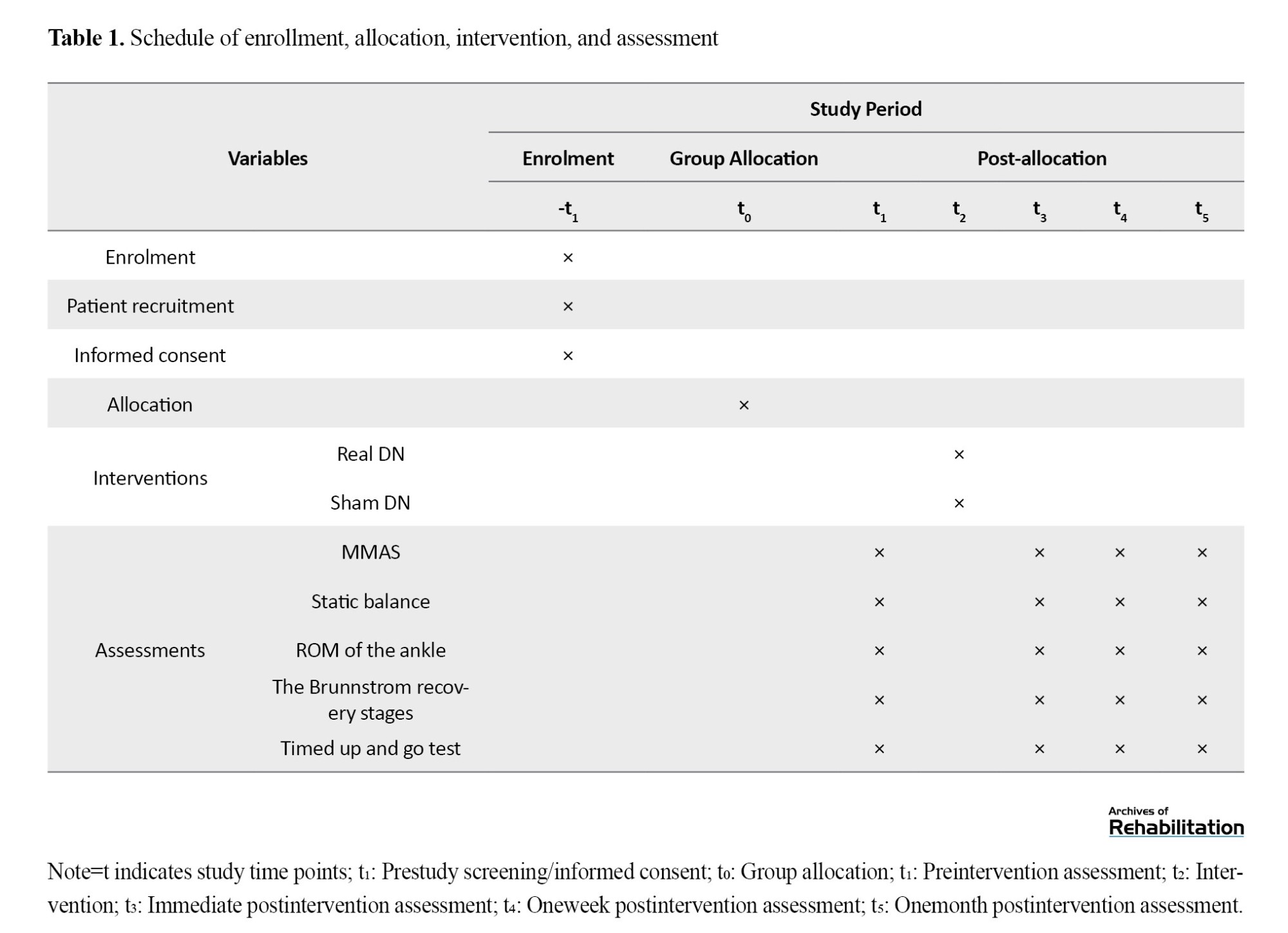
This is a protocol study for a randomized, double-blind, parallel-group clinical trial, designed according to the standard protocol items: recommendations for Interventional Trials (SPIRIT) guidelines [17] as shown in Table 1.
Study area, participants, sampling, and allocation
Participants will be patients with stroke. At their first visit to the Musculoskeletal Research Center, School of Rehabilitation Sciences, Isfahan University of Medical Sciences, Isfahan, Iran, they will undergo a comprehensive initial assessment and, based on the study inclusion and exclusion criteria, those deemed eligible will be selected and entered into subsequent stages of the research. All phases of the trial, including assessments, treatment, and follow-up, will be conducted at the aforementioned research center.
The first session is expected to last approximately 1.5 hours due to the completion of the informed consent form and patient information forms, whereas the follow-up sessions (one week and one month post-intervention) each will take about 1 hour. After each visit, the patient will be offered light refreshments and, following confirmation of their general well-being, will be discharged.
The entry criteria will include: diagnosis of stroke, age 45-75 years, at least 6 months have passed since the onset of stroke, spasticity intensity of the plantar flexor and inverter muscles ≥1 based on the MMAS score, and ability to stand and walk independently. Exclusion criteria will be the use of anti-spasticity medications, presence of other neurological disorders, history of fracture or surgery in lower limbs, presence of a fixed deformity in the affected ankle, contraindications to DN (e.g. needle phobia), participation in other clinical trials, and unwillingness to participate.
Significance level (α)=0.05, test power (1-β)=85%, effect size (Δ)=1 unit for the mean difference between the two groups, standard deviation of 0.78 for the intervention group and 0.89 for the sham group, the allocation ratio 1:1. Applying these assumptions to the formula and using the values (Z(1-β)=1.04) and (Z(1-α/2)=1.96), the required sample size was calculated to be 13 per group. Accordingly, the total required sample size was 26 (Equation 1).
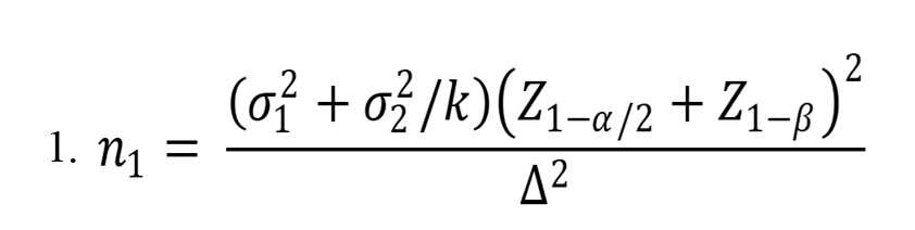
Where, n1=calculated sample size for group A=13; n2=calculated sample size for group B=13; σ1=standard deviation of group A=0.78; σ2=standard deviation of group B=0.89; Δ=Mean difference between the two groups=1; k=n2/n1=1; Z1- α/2=two-tailed z value (1.96 for 95% confidence interval); Z1-β=statistical power=85%.
To recruit patients, in addition to consulting and requesting neurologists in Isfahan, we will contact with hospitals and specialized treatment centers, including Al-Zahra, Amin, Shariati, and Gharazi hospitals in Isfahan. Sampling will be conducted using a convenience method. Allocation of patients to the intervention and sham groups will be performed using the block randomization method (block size of 4, allocation ratio 1:1) using a random number generator to ensure an equal distribution of patients in each group [16]. To conceal the randomization sequence, sequentially numbered, sealed, opaque envelopes containing letters A or B will be used. Patients in group A will receive real DN on the gastrocnemius, soleus, and tibialis posterior muscles, while patients in group B will receive sham DN on the same muscles. The research center secretary, who will be provided with the envelopes, will not be involved in any assessment or treatment stages. The physiotherapist will receive the sealed envelopes and will administer the treatment accordingly. The non-treating physiotherapist (assessor) and the patients will remain blinded to the type of treatment.
Intervention
Individuals in the intervention group A will undergo a single DN session targeting the approximate motor points of the gastrocnemius, soleus, and tibialis posterior muscles. For DN, sterile, single-use, stainless steel acupuncture needles measuring 0.30 mm × 50 mm (Dong Bang AcuPrime, South Korea) will be used. Prior to needle insertion, the patient’s skin will be disinfected with an alcohol swab. Using a fast-in, fast-out pistoning technique, the needle will be inserted to the motor points of the muscles and will be removed for 1 minute [7]. After needle removal, the site will be compressed for 5 seconds and then cleaned again with an alcohol swab. Used needles will be disposed of in a designated sharps container.
The muscles selected for DN were as follows:
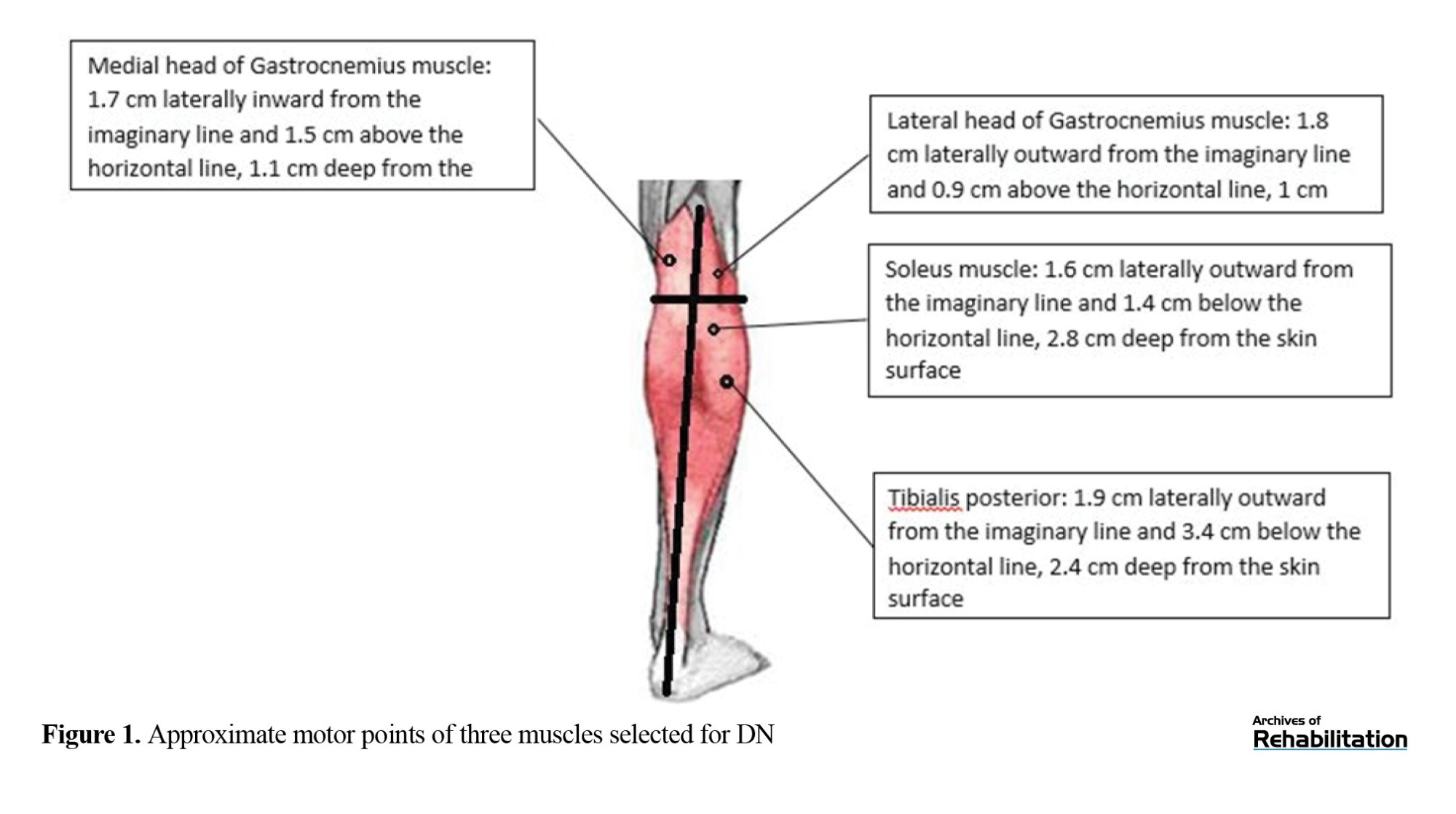
Medial gastrocnemius muscle: In a prone position, a pillow under the patient’s tibial crest and the ankle in plantar flexion, the needle will be inserted in the vertical axis 1.5 cm above the horizontal line and 1.7 cm in the horizontal axis medial to the assumed midline of the popliteal fossa (Figure 1).
Lateral gastrocnemius muscle: In the same position, the needle will be inserted 0.9 cm above the horizontal line and 1.8 cm lateral to the assumed midline of the popliteal fossa, at a depth of 1 cm from the skin surface (Figure 1).
Soleus muscle: In the prone position and the knee extended, the needle will be inserted 1.4 cm inferior to the horizontal line and 1.6 cm lateral to the assumed midline (Figure 1).
Tibialis posterior muscle: In the same position, the needle will be inserted 4.3 cm inferior to the horizontal line and 1.9 cm lateral to the assumed midline (Figure 1) [10].
Primary outcomes
Spasticity
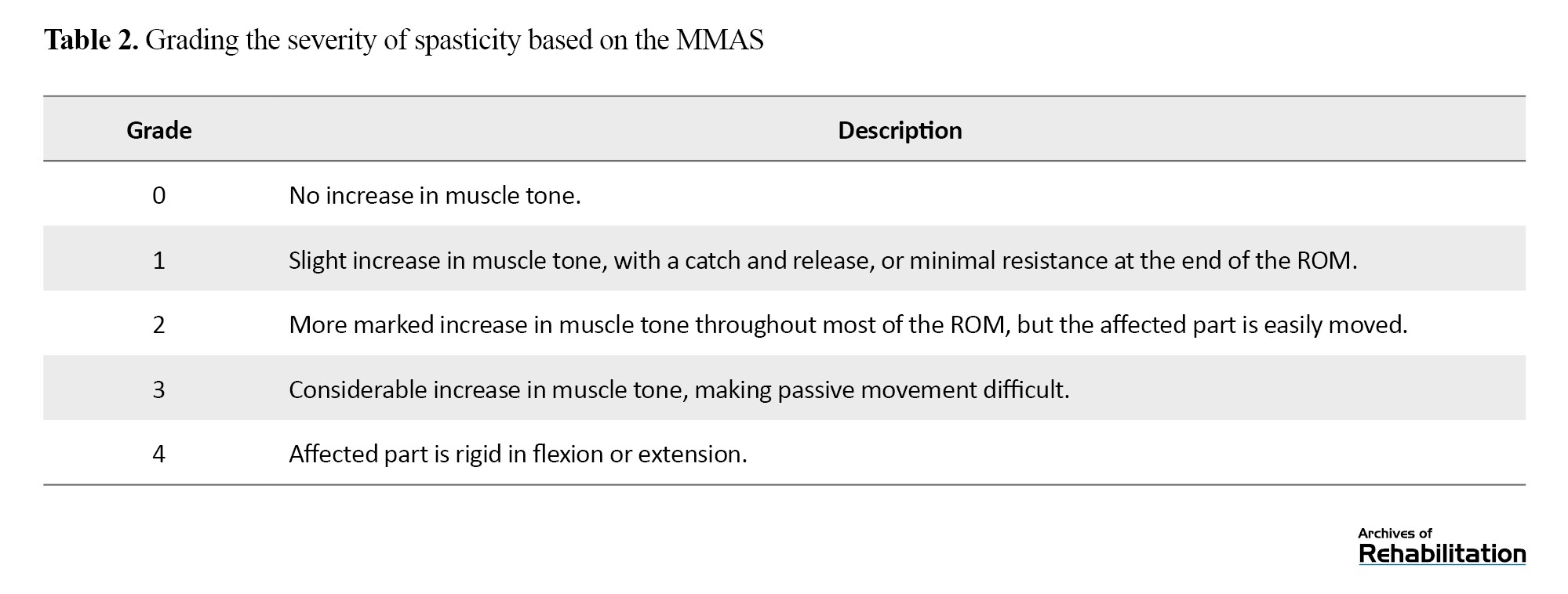
Spasticity of the ankle plantar-flexor muscles will be evaluated using the Persian version of the MMAS [11]. It grades the severity of spasticity according to the degree of resistance to passive movement, ranging from grade 0 (no increase in muscle tone) to grade 4 (rigi dity of the affected parts in flexion or extension) (Table 2). The inter-rater and intra-rater reliability of the Persian MMAS for assessing lower limb muscles in stroke patients has been confirmed [12]. A passive movement will be used to grade the severity of spasticity. To reduce dependence on the examiner’s judgment and skill, this assessment will be conducted under the supervision of assessors with prior experience in spasticity evaluation.
Static balance
Static balance will be assessed using the posturography method with a Kistler force plate (Model 5233 A2, Kistler Instrumente AG, Switzerland). The use of force plates for measuring balance in stroke patients has been validated in previous studies [13]. In this test, the physiotherapist will instruct the patient to stand barefoot on the force plate with heels 9 cm apart, feet at a 30-degree angle, and upper limbs relaxed at the sides. Under eyes-open conditions, the patient is asked to fixate on a point on the wall at a distance of 2 meters. Under eyes-closed conditions, the test will be performed without visual fixation. The sequence of eyes-open and eyes-closed conditions will be randomized, with a 2-minute rest interval between them. Each will be performed three times, with a 20-second interval between repetitions. The duration of each trial will be 20 seconds. Velocity (cm/s) and anteroposterior and mediolateral center of pressure (COP) displacements will be recorded across the three trials, and their averages will be calculated for analysis [18].
Secondary outcomes
Ankle range of motion
Passive ankle ROM will be measured in degrees using a standard goniometer in supine position with the knee extended. The neutral position is considered to be the ankle joint at a 90-degree angle. For ROM assessment, the goniometer’s axis will be placed over the lateral malleolus, the stationary arm will be aligned with the fibular head, and the movable arm will be aligned with the fifth metatarsal. The physiotherapist will stabilize the affected foot with one hand and move the ankle into maximal passive dorsiflexion with the other hand. For assessing active ROM, the same procedure will be used, except the patient will be instructed to actively move their ankle [14].
Lower limb function
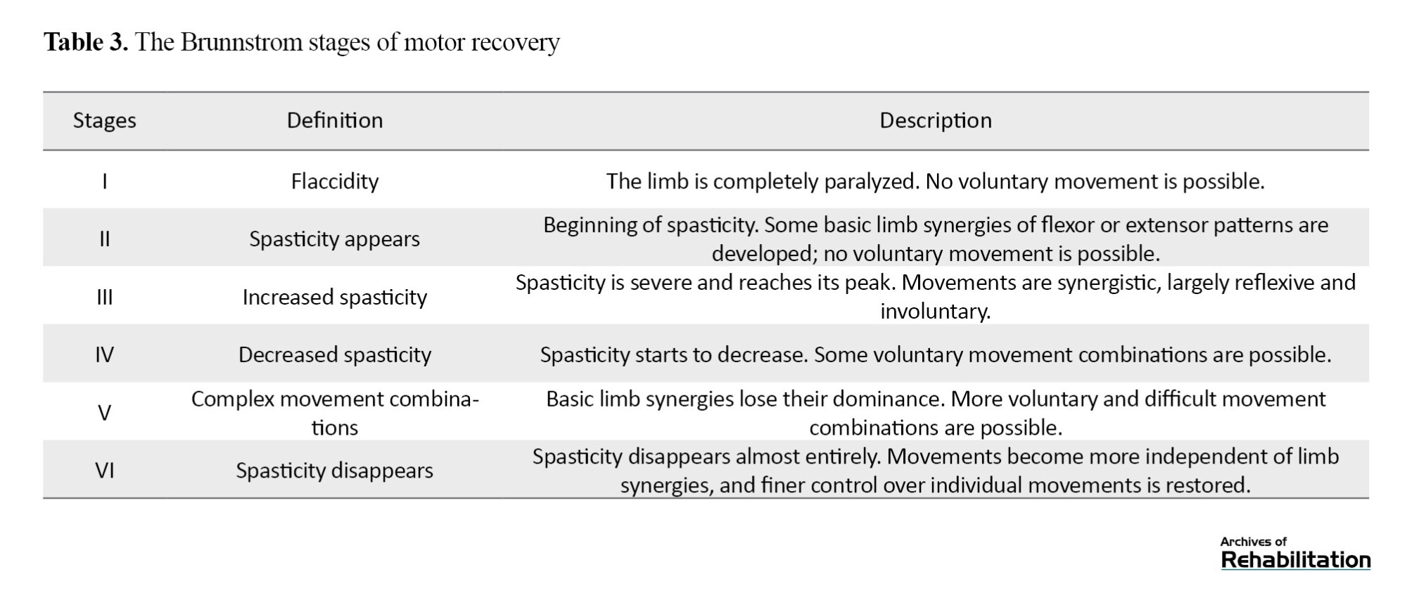
The lower-limb motor recovery process will be assessed using the Brunnstrom scale (Table 3). This method classifies patient recovery into six stages, from stage 1 (flaccidity) to stage 6 (disappearance of spasticity) [15].
Dynamic balance
Dynamic balance and functional mobility will be measured using the timed up and go (TUG) test, in which the participant rises from a chair, walks three meters, turns, returns to the chair, and sits down. The total time taken is recorded as the test score. This test is recognized as a valid tool for assessing mobility and dynamic balance in various conditions, including stroke [19].
Data and adverse effect monitoring
An independent panel of experts (faculty members from various rehabilitation fields) will supervise the study procedures to ensure methodological accuracy. This supervision includes not only adherence to the proposed protocol but also guarantees the accuracy of data collection. This expert panel will help in maintaining data reliability and ultimately contribute to optimizing the results and progress of the study.
Throughout the study, observed adverse effects will be monitored and recorded. In case of any adverse effect, standard medical care will be promptly provided, and the patient’s participation in the study will be discontinued. This precaution is taken to ensure patient safety and well-being.
Statistical analysis
Data analysis will be conducted in SPSS software, version 24, using descriptive statistics such as Mean±SD, median, and interquartile range. Normality will be assessed using the Kolmogorov–Smirnov test. The homogeneity of variances between groups will be assessed using Levene’s test. Depending on the normality/abnormality of the data distribution, baseline comparisons between groups will be carried out using an independent t-test or the Mann–Whitney U test. If the outcome measures were normally distributed, a two-way repeated measures analysis of variance (ANOVA) will be employed to examine the effects of “time”, “group”, and the “time×group” interaction. The Bonferroni correction will be applied for pairwise comparisons between time points. Subsequently, a generalized estimating equation model was used for the analysis of repeated-measures data. This model was applied to compare changes in the MMAS score between the groups and over time. Effect size was calculated using Cohen’s d and interpreted as follows:
Trivial: <0.20;
Small: 0.20-0.50;
Medium: 0.50-0.80;
Large: ≥0.80.
Discussion
The present study provided the protocol of a clinical trial to compare the effects of real DN versus sham DN on spasticity of the ankle plantar flexor and invertor muscles, as well as balance impairments, in patients with stroke. Few high-quality studies and randomized clinical trials are available on the effectiveness of DN in improving ankle spasticity and balance after stroke [5]. A key feature of the present protocol is its randomized and parallel allocation of patients to treatment groups. This methodology is intended to reduce confounding variables and bias, thereby enhancing the reliability and validity of the study findings [16]. Ensuring that both patients and outcome assessors are blinded to treatment allocation reduces the risk of bias in treatment allocation and outcome evaluation, thereby strengthening the study findings.
Our hypothesis is that real DN will be more effective than sham DN in reducing spasticity and balance impairment following stroke. Based on prior evidence and our clinical expertise, a reduction in spasticity and improvement in static and dynamic balance may be observed in stroke patients receiving real DN compared with those receiving sham DN [4, 8]. Ebrahimi et al. showed that DN combined with exercise, compared to exercise alone, improved spasticity, ROM, and function in patients with chronic post-stroke spasticity [20]. Given the limitations reported in previous studies regarding clinically meaningful improvements in active ankle ROM, achieving improvement in this type of ROM may be challenging [21].
The probable beneficial therapeutic effects of real DN in addressing certain problems in patients with stroke can be associated with improvements in ankle spasticity, which may be due to nervous system involvement following stroke [21]. Indeed, both neural and peripheral mechanisms may be involved in the development of spasticity [21, 22]. An imbalance in the descending supraspinal pathways may lead to dysregulation of the stretch reflex and hyperexcitability of alpha motor neurons [21]. In addition to the potential benefits reported for the treatment of spasticity with DN, some studies have also emphasized the safety of this intervention [23]. This, combined with structural changes in muscles affected after stroke, causes muscle contractions and stiffness. Pain is commonly observed in stroke patients with spasticity and worsens their condition. Structural changes in spastic muscles and shared central neural networks for pain and spasticity have been reported [22, 24]. Therefore, it is hypothesized that both neurophysiological mechanisms (such as modulation of alpha motor neuron hyperexcitability, activation of the inhibitory system, and pain reduction) and mechanical effects, including localized stretching of cytoskeletal elements and reduced overlap between actin and myosin filaments, may contribute to the observed reductions in spasticity following real DN [22, 24].
The probable improvement in ankle ROM and balance after DN may be attributed to various factors. First, the reduction in spasticity and muscle stiffness, documented in previous studies, can provide greater freedom for ankle joint movement and increase ROM [8]. This reduction in muscle tone, in turn, may facilitate improved balance function [8]. Furthermore, by reducing spasticity and enhancing brain activity, real DN may contribute to more stable and controlled movement, thereby improving active ankle ROM [4, 8]. In addition, increased support for the affected limb, resulting from reduced muscle stiffness and spasticity, may enhance proprioceptive feedback and stability, thereby improving balance outcomes [8, 13]. Overall, these combined effects indicate the multifaceted benefits of real DN in treating spasticity as well as its broader impact on motor function and balance in stroke patients.
The clinical trial will determine how DN influences spasticity and balance deficits in individuals after stroke. Given existing evidence that this method significantly reduces muscle spasticity and improves balance indices [4, 5], the results of this study are expected to align with these findings. In examining discrepancies among studies, it should be noted that one study has not confirmed a significant effect of DN on spasticity [5], while a study reported positive effects of DN even in short-term interventions [4]. Other studies, employing longer follow-up periods, have reported findings consistent with the present study’s expected outcomes and emphasized that DN can be a useful complement to conventional rehabilitation therapies after stroke [5, 8].
A notable advantage of the clinical trial will be the use of validated instruments to evaluate spasticity and balance, which can enhance the reliability and generalizability of its outcomes. The results of this study will likely have significant clinical applications. For example, adding DN to conventional rehabilitation protocols, as shown in studies such as Sánchez Milá et al.’s study [4], may accelerate motor function improvement and reduce disability in patients by rapidly reducing spasticity and improving balance. Recent systematic reviews also emphasize that despite observing reduced spasticity post-intervention in some studies, a significant impact on balance and motor function in stroke patients has not been conclusively demonstrated, and some studies, due to lack of long-term follow-up or problems in study design, have been unable to prove definitive efficacy [5]. Differences in study results can be attributed to factors such as sample heterogeneity (age, time since stroke onset, spasticity severity), differences in intervention protocol and duration, target muscles, and assessment tools [5].
Conclusion
Based on existing evidence from prior studies, it is predicted that the results of the clinical trial will show short-term reduction in spasticity and occasional improvement in motor function in patients with stroke after DN. Its combined approach, which simultaneously assesses several key outcomes and conducts follow-up at multiple time points, is likely to address some of the gaps identified in previous studies.
The clinical trial, however, will have some limitations. Despite employing bias-reduction strategies such as randomization and blinding, some uncontrollable factors, including individual differences and environmental variables, may affect the results. Furthermore, the relatively short duration of the treatment and follow-up period may prevent us from capturing the full spectrum of therapeutic effects over a longer period. Therefore, future research should include long-term follow-up periods to better understand the lasting effects of DN over time.
Ethical Considerations
Compliance with ethical guidelines
The proposal of this study was approved by the Ethics Committee of the University of Social Welfare and Rehabilitation Sciences (Code: IR.USWR.REC.1401.189). This research is a protocol for a randomized clinical trial, registered by the Iranian Registry of Clinical Trials (ID: IRCT20221228056963N1). Written informed consent will be obtained from patients prior to their participation. All procedures will be conducted in accordance with the Declaration of Helsinki.
Funding
This article was extracted from the doctoral thesis of Seyed Mehdi Okhravi at the Department of Physiotherapy, University of Social Welfare and Rehabilitation Sciences. This research did not receive any grant from funding agencies in the public, commercial, or non-profit sectors.
Authors' contributions
Conceptualization, methodology, and writing: Noureddin Nakhostin Ansari, Iraj Abdollahi, and Seyed Mehdi Okhravi; Investigation, resources, and funding acquisition: Seyed Mehdi Okhravi; Translation and editing: Seyed Mehdi Okhravi and Seyed Mehdi Mohsenipour.
Conflict of interest
The authors declare no conflict of interests.
AI tools disclosure statement
The authors used ChatGPT solely to improve the readability, language, and overall quality of writing. All authors remain fully responsible for scientific content, intervention protocols, statistical analyses, interpretations, and conclusions.
Acknowledgments
The authors would like to thank all individuals who will participate in this research.
Stroke remains a major global concern that contributes to long-term disability and is associated with neuromuscular issues such as increased muscle tone, impaired balance, diminished range of motion, and reduced functional mobility [1]. In recent years, dry needling (DN) has attracted researchers’ attention as a minimally invasive adjunctive technique in physiotherapy to improve functional outcomes in patients with stroke [2]. However, a review of the literature indicates that previous studies have mainly focused on the upper extremity or on specific muscles such as the gastrocnemius or tibialis anterior, with less attention given to the simultaneous study of multiple key muscles of the lower extremity [3, 4]. The disparities in findings regarding the effectiveness of DN for spasticity are another limitation [5]. These differences in findings may be attributed to variability in the intervention protocols, assessment tools, target muscles, patient characteristics, and follow-up duration [6]. On the other hand, the precise mechanism of action of DN has not yet been fully elucidated, and considerable uncertainty remains regarding the role of neurophysiological factors (such as reduced alpha motor neuron excitability and activation of central inhibitory pathways) as well as mechanical factors (such as the release of myofascial trigger points or improvement in local blood flow) [5].
As mentioned above, one of the main limitations of previous studies is the failure to investigate simultaneously the effects of DN on multiple key muscles involved in lower-limb spasticity. The spastic gait pattern often requires a more comprehensive approach that accounts for the kinetic chain and the roles of different muscles. Most previous studies have examined only one of these muscles or used a single, restricted method of functional assessment [5, 7, 8]. This point is important because, in most cases, lower-limb spasticity in patients with stroke involves not a single muscle but multiple muscles, and the interaction among these muscles may influence treatment outcomes. Previous studies have largely focused on a single muscle, such as the gastrocnemius, whereas the spastic gait pattern often requires a more comprehensive approach that considers the kinetic chain and the role of different muscles within it. This multi-muscle approach more accurately reflects the patients’ actual clinical presentation and may promote greater overall functional improvement. Many studies address only one or two outcomes. For example, some have focused solely on reducing spasticity, neglecting to evaluate its effects on balance or overall function [5]. Moreover, many previous studies have lacked a follow-up period [6, 9]. For instance, Mendigutía-Gómez et al. primarily focused on immediate and short-term effects [5]. This is particularly important because the short-term effectiveness of an intervention might arise from various factors, including placebo effects or temporary physiological changes. What is particularly important in rehabilitation interventions is the extent to which therapeutic effects remain stable and durable over time. Moreover, most studies have lacked a double-blind design and a placebo group, which may reduce the internal validity of their findings.
The present study provides the protocol of a clinical trial to simultaneously investigate the effects of DN on three key lower limb muscles including the gastrocnemius, soleus, and tibialis posterior, in patients with stroke. Another innovation of this study will be the comprehensive assessment of several key outcomes: spasticity using the modified Ashworth scale (MMAS) [10-12], balance via posturography [13] range of motion (ROM) using a goniometer [14], and overall lower limb function based on the Brunnstrom stages of stroke recovery [15]. This integrated approach aims to investigate the potential cause-and-effect relationships between these outcomes, yielding a deeper understanding of recovery mechanisms. By utilizing valid and standard tools such as the MMAS for spasticity, posturography for quantitative balance assessment (including advanced indices such as center of pressure sway area and center of pressure displacement velocity), a goniometer for measuring ROM, and the Brunnstrom scale for comprehensive motor function evaluation, this study will likely provide a holistic picture of the DN effects. Moreover, the use of follow-up periods for assessing outcomes (immediately post-intervention, one week post-intervention, and one month post-intervention) allows for a more accurate measurement of the intervention’s effect sustainability. The follow-up assessments enable us to better understand the persistence of DN’s therapeutic effects. Clarifying this will likely improve understanding of future treatment protocols. Another strength of the clinical trial will be the use of double-blinding (where both patients and assessors are blind to the intervention type), a sham placebo group, and a block randomization method, which can enhance the validity of the results [16].
These are fundamental pillars of high-quality clinical trials. Double-blinding minimizes bias arising from expectations. The sham intervention, which involves a similar but therapeutically inactive method (e.g. superficial DN without penetrating the muscle motor point), enables the differentiation of genuine therapeutic effects from non-specific placebo effects. Block randomization can maintain a balanced distribution of baseline variables such as age, sex, and stroke severity between the intervention and sham groups.
Therefore, the clinical trial will prove the efficacy of DN, will provide clinical evidence regarding the multi-faceted relationships between major stroke rehabilitation outcomes, and will determine the sustainability of DN effects. Such evidence can offer a new and practical model for evidence-based rehabilitation protocols. The clinical trial goes beyond a simple “efficacy study” and attempts to position DN as an important functional therapy for the treatment of patients with spasticity. The results can help physicians, physiotherapists, and other rehabilitation specialists develop and implement more comprehensive, evidence-based treatment protocols to improve the quality of life for stroke patients.
Materials and Methods
Study design
This is a protocol study for a randomized, double-blind, parallel-group clinical trial, designed according to the standard protocol items: recommendations for Interventional Trials (SPIRIT) guidelines [17] as shown in Table 1.
Study area, participants, sampling, and allocation
Participants will be patients with stroke. At their first visit to the Musculoskeletal Research Center, School of Rehabilitation Sciences, Isfahan University of Medical Sciences, Isfahan, Iran, they will undergo a comprehensive initial assessment and, based on the study inclusion and exclusion criteria, those deemed eligible will be selected and entered into subsequent stages of the research. All phases of the trial, including assessments, treatment, and follow-up, will be conducted at the aforementioned research center.
The first session is expected to last approximately 1.5 hours due to the completion of the informed consent form and patient information forms, whereas the follow-up sessions (one week and one month post-intervention) each will take about 1 hour. After each visit, the patient will be offered light refreshments and, following confirmation of their general well-being, will be discharged.
The entry criteria will include: diagnosis of stroke, age 45-75 years, at least 6 months have passed since the onset of stroke, spasticity intensity of the plantar flexor and inverter muscles ≥1 based on the MMAS score, and ability to stand and walk independently. Exclusion criteria will be the use of anti-spasticity medications, presence of other neurological disorders, history of fracture or surgery in lower limbs, presence of a fixed deformity in the affected ankle, contraindications to DN (e.g. needle phobia), participation in other clinical trials, and unwillingness to participate.
Significance level (α)=0.05, test power (1-β)=85%, effect size (Δ)=1 unit for the mean difference between the two groups, standard deviation of 0.78 for the intervention group and 0.89 for the sham group, the allocation ratio 1:1. Applying these assumptions to the formula and using the values (Z(1-β)=1.04) and (Z(1-α/2)=1.96), the required sample size was calculated to be 13 per group. Accordingly, the total required sample size was 26 (Equation 1).
Where, n1=calculated sample size for group A=13; n2=calculated sample size for group B=13; σ1=standard deviation of group A=0.78; σ2=standard deviation of group B=0.89; Δ=Mean difference between the two groups=1; k=n2/n1=1; Z1- α/2=two-tailed z value (1.96 for 95% confidence interval); Z1-β=statistical power=85%.
To recruit patients, in addition to consulting and requesting neurologists in Isfahan, we will contact with hospitals and specialized treatment centers, including Al-Zahra, Amin, Shariati, and Gharazi hospitals in Isfahan. Sampling will be conducted using a convenience method. Allocation of patients to the intervention and sham groups will be performed using the block randomization method (block size of 4, allocation ratio 1:1) using a random number generator to ensure an equal distribution of patients in each group [16]. To conceal the randomization sequence, sequentially numbered, sealed, opaque envelopes containing letters A or B will be used. Patients in group A will receive real DN on the gastrocnemius, soleus, and tibialis posterior muscles, while patients in group B will receive sham DN on the same muscles. The research center secretary, who will be provided with the envelopes, will not be involved in any assessment or treatment stages. The physiotherapist will receive the sealed envelopes and will administer the treatment accordingly. The non-treating physiotherapist (assessor) and the patients will remain blinded to the type of treatment.
Intervention
Individuals in the intervention group A will undergo a single DN session targeting the approximate motor points of the gastrocnemius, soleus, and tibialis posterior muscles. For DN, sterile, single-use, stainless steel acupuncture needles measuring 0.30 mm × 50 mm (Dong Bang AcuPrime, South Korea) will be used. Prior to needle insertion, the patient’s skin will be disinfected with an alcohol swab. Using a fast-in, fast-out pistoning technique, the needle will be inserted to the motor points of the muscles and will be removed for 1 minute [7]. After needle removal, the site will be compressed for 5 seconds and then cleaned again with an alcohol swab. Used needles will be disposed of in a designated sharps container.
The muscles selected for DN were as follows:
Medial gastrocnemius muscle: In a prone position, a pillow under the patient’s tibial crest and the ankle in plantar flexion, the needle will be inserted in the vertical axis 1.5 cm above the horizontal line and 1.7 cm in the horizontal axis medial to the assumed midline of the popliteal fossa (Figure 1).
Lateral gastrocnemius muscle: In the same position, the needle will be inserted 0.9 cm above the horizontal line and 1.8 cm lateral to the assumed midline of the popliteal fossa, at a depth of 1 cm from the skin surface (Figure 1).
Soleus muscle: In the prone position and the knee extended, the needle will be inserted 1.4 cm inferior to the horizontal line and 1.6 cm lateral to the assumed midline (Figure 1).
Tibialis posterior muscle: In the same position, the needle will be inserted 4.3 cm inferior to the horizontal line and 1.9 cm lateral to the assumed midline (Figure 1) [10].
Primary outcomes
Spasticity
Spasticity of the ankle plantar-flexor muscles will be evaluated using the Persian version of the MMAS [11]. It grades the severity of spasticity according to the degree of resistance to passive movement, ranging from grade 0 (no increase in muscle tone) to grade 4 (rigi dity of the affected parts in flexion or extension) (Table 2). The inter-rater and intra-rater reliability of the Persian MMAS for assessing lower limb muscles in stroke patients has been confirmed [12]. A passive movement will be used to grade the severity of spasticity. To reduce dependence on the examiner’s judgment and skill, this assessment will be conducted under the supervision of assessors with prior experience in spasticity evaluation.
Static balance
Static balance will be assessed using the posturography method with a Kistler force plate (Model 5233 A2, Kistler Instrumente AG, Switzerland). The use of force plates for measuring balance in stroke patients has been validated in previous studies [13]. In this test, the physiotherapist will instruct the patient to stand barefoot on the force plate with heels 9 cm apart, feet at a 30-degree angle, and upper limbs relaxed at the sides. Under eyes-open conditions, the patient is asked to fixate on a point on the wall at a distance of 2 meters. Under eyes-closed conditions, the test will be performed without visual fixation. The sequence of eyes-open and eyes-closed conditions will be randomized, with a 2-minute rest interval between them. Each will be performed three times, with a 20-second interval between repetitions. The duration of each trial will be 20 seconds. Velocity (cm/s) and anteroposterior and mediolateral center of pressure (COP) displacements will be recorded across the three trials, and their averages will be calculated for analysis [18].
Secondary outcomes
Ankle range of motion
Passive ankle ROM will be measured in degrees using a standard goniometer in supine position with the knee extended. The neutral position is considered to be the ankle joint at a 90-degree angle. For ROM assessment, the goniometer’s axis will be placed over the lateral malleolus, the stationary arm will be aligned with the fibular head, and the movable arm will be aligned with the fifth metatarsal. The physiotherapist will stabilize the affected foot with one hand and move the ankle into maximal passive dorsiflexion with the other hand. For assessing active ROM, the same procedure will be used, except the patient will be instructed to actively move their ankle [14].
Lower limb function
The lower-limb motor recovery process will be assessed using the Brunnstrom scale (Table 3). This method classifies patient recovery into six stages, from stage 1 (flaccidity) to stage 6 (disappearance of spasticity) [15].
Dynamic balance
Dynamic balance and functional mobility will be measured using the timed up and go (TUG) test, in which the participant rises from a chair, walks three meters, turns, returns to the chair, and sits down. The total time taken is recorded as the test score. This test is recognized as a valid tool for assessing mobility and dynamic balance in various conditions, including stroke [19].
Data and adverse effect monitoring
An independent panel of experts (faculty members from various rehabilitation fields) will supervise the study procedures to ensure methodological accuracy. This supervision includes not only adherence to the proposed protocol but also guarantees the accuracy of data collection. This expert panel will help in maintaining data reliability and ultimately contribute to optimizing the results and progress of the study.
Throughout the study, observed adverse effects will be monitored and recorded. In case of any adverse effect, standard medical care will be promptly provided, and the patient’s participation in the study will be discontinued. This precaution is taken to ensure patient safety and well-being.
Statistical analysis
Data analysis will be conducted in SPSS software, version 24, using descriptive statistics such as Mean±SD, median, and interquartile range. Normality will be assessed using the Kolmogorov–Smirnov test. The homogeneity of variances between groups will be assessed using Levene’s test. Depending on the normality/abnormality of the data distribution, baseline comparisons between groups will be carried out using an independent t-test or the Mann–Whitney U test. If the outcome measures were normally distributed, a two-way repeated measures analysis of variance (ANOVA) will be employed to examine the effects of “time”, “group”, and the “time×group” interaction. The Bonferroni correction will be applied for pairwise comparisons between time points. Subsequently, a generalized estimating equation model was used for the analysis of repeated-measures data. This model was applied to compare changes in the MMAS score between the groups and over time. Effect size was calculated using Cohen’s d and interpreted as follows:
Trivial: <0.20;
Small: 0.20-0.50;
Medium: 0.50-0.80;
Large: ≥0.80.
Discussion
The present study provided the protocol of a clinical trial to compare the effects of real DN versus sham DN on spasticity of the ankle plantar flexor and invertor muscles, as well as balance impairments, in patients with stroke. Few high-quality studies and randomized clinical trials are available on the effectiveness of DN in improving ankle spasticity and balance after stroke [5]. A key feature of the present protocol is its randomized and parallel allocation of patients to treatment groups. This methodology is intended to reduce confounding variables and bias, thereby enhancing the reliability and validity of the study findings [16]. Ensuring that both patients and outcome assessors are blinded to treatment allocation reduces the risk of bias in treatment allocation and outcome evaluation, thereby strengthening the study findings.
Our hypothesis is that real DN will be more effective than sham DN in reducing spasticity and balance impairment following stroke. Based on prior evidence and our clinical expertise, a reduction in spasticity and improvement in static and dynamic balance may be observed in stroke patients receiving real DN compared with those receiving sham DN [4, 8]. Ebrahimi et al. showed that DN combined with exercise, compared to exercise alone, improved spasticity, ROM, and function in patients with chronic post-stroke spasticity [20]. Given the limitations reported in previous studies regarding clinically meaningful improvements in active ankle ROM, achieving improvement in this type of ROM may be challenging [21].
The probable beneficial therapeutic effects of real DN in addressing certain problems in patients with stroke can be associated with improvements in ankle spasticity, which may be due to nervous system involvement following stroke [21]. Indeed, both neural and peripheral mechanisms may be involved in the development of spasticity [21, 22]. An imbalance in the descending supraspinal pathways may lead to dysregulation of the stretch reflex and hyperexcitability of alpha motor neurons [21]. In addition to the potential benefits reported for the treatment of spasticity with DN, some studies have also emphasized the safety of this intervention [23]. This, combined with structural changes in muscles affected after stroke, causes muscle contractions and stiffness. Pain is commonly observed in stroke patients with spasticity and worsens their condition. Structural changes in spastic muscles and shared central neural networks for pain and spasticity have been reported [22, 24]. Therefore, it is hypothesized that both neurophysiological mechanisms (such as modulation of alpha motor neuron hyperexcitability, activation of the inhibitory system, and pain reduction) and mechanical effects, including localized stretching of cytoskeletal elements and reduced overlap between actin and myosin filaments, may contribute to the observed reductions in spasticity following real DN [22, 24].
The probable improvement in ankle ROM and balance after DN may be attributed to various factors. First, the reduction in spasticity and muscle stiffness, documented in previous studies, can provide greater freedom for ankle joint movement and increase ROM [8]. This reduction in muscle tone, in turn, may facilitate improved balance function [8]. Furthermore, by reducing spasticity and enhancing brain activity, real DN may contribute to more stable and controlled movement, thereby improving active ankle ROM [4, 8]. In addition, increased support for the affected limb, resulting from reduced muscle stiffness and spasticity, may enhance proprioceptive feedback and stability, thereby improving balance outcomes [8, 13]. Overall, these combined effects indicate the multifaceted benefits of real DN in treating spasticity as well as its broader impact on motor function and balance in stroke patients.
The clinical trial will determine how DN influences spasticity and balance deficits in individuals after stroke. Given existing evidence that this method significantly reduces muscle spasticity and improves balance indices [4, 5], the results of this study are expected to align with these findings. In examining discrepancies among studies, it should be noted that one study has not confirmed a significant effect of DN on spasticity [5], while a study reported positive effects of DN even in short-term interventions [4]. Other studies, employing longer follow-up periods, have reported findings consistent with the present study’s expected outcomes and emphasized that DN can be a useful complement to conventional rehabilitation therapies after stroke [5, 8].
A notable advantage of the clinical trial will be the use of validated instruments to evaluate spasticity and balance, which can enhance the reliability and generalizability of its outcomes. The results of this study will likely have significant clinical applications. For example, adding DN to conventional rehabilitation protocols, as shown in studies such as Sánchez Milá et al.’s study [4], may accelerate motor function improvement and reduce disability in patients by rapidly reducing spasticity and improving balance. Recent systematic reviews also emphasize that despite observing reduced spasticity post-intervention in some studies, a significant impact on balance and motor function in stroke patients has not been conclusively demonstrated, and some studies, due to lack of long-term follow-up or problems in study design, have been unable to prove definitive efficacy [5]. Differences in study results can be attributed to factors such as sample heterogeneity (age, time since stroke onset, spasticity severity), differences in intervention protocol and duration, target muscles, and assessment tools [5].
Conclusion
Based on existing evidence from prior studies, it is predicted that the results of the clinical trial will show short-term reduction in spasticity and occasional improvement in motor function in patients with stroke after DN. Its combined approach, which simultaneously assesses several key outcomes and conducts follow-up at multiple time points, is likely to address some of the gaps identified in previous studies.
The clinical trial, however, will have some limitations. Despite employing bias-reduction strategies such as randomization and blinding, some uncontrollable factors, including individual differences and environmental variables, may affect the results. Furthermore, the relatively short duration of the treatment and follow-up period may prevent us from capturing the full spectrum of therapeutic effects over a longer period. Therefore, future research should include long-term follow-up periods to better understand the lasting effects of DN over time.
Ethical Considerations
Compliance with ethical guidelines
The proposal of this study was approved by the Ethics Committee of the University of Social Welfare and Rehabilitation Sciences (Code: IR.USWR.REC.1401.189). This research is a protocol for a randomized clinical trial, registered by the Iranian Registry of Clinical Trials (ID: IRCT20221228056963N1). Written informed consent will be obtained from patients prior to their participation. All procedures will be conducted in accordance with the Declaration of Helsinki.
Funding
This article was extracted from the doctoral thesis of Seyed Mehdi Okhravi at the Department of Physiotherapy, University of Social Welfare and Rehabilitation Sciences. This research did not receive any grant from funding agencies in the public, commercial, or non-profit sectors.
Authors' contributions
Conceptualization, methodology, and writing: Noureddin Nakhostin Ansari, Iraj Abdollahi, and Seyed Mehdi Okhravi; Investigation, resources, and funding acquisition: Seyed Mehdi Okhravi; Translation and editing: Seyed Mehdi Okhravi and Seyed Mehdi Mohsenipour.
Conflict of interest
The authors declare no conflict of interests.
AI tools disclosure statement
The authors used ChatGPT solely to improve the readability, language, and overall quality of writing. All authors remain fully responsible for scientific content, intervention protocols, statistical analyses, interpretations, and conclusions.
Acknowledgments
The authors would like to thank all individuals who will participate in this research.
References:
- GBD 2019 Stroke Collaborators. Global, regional, and national burden of stroke and its risk factors, 1990-2019: A systematic analysis for the Global Burden of Disease Study 2019. The Lancet. Neurology. 2021; 20(10):795-820. [DOI:10.1016/S1474-4422(21)00252-0] [PMID]
- Zhang Z, Wang W, Song Y, Zhai T, Zhu Y, Jiang L, et al. Immediate effect of dry needling at myofascial trigger point on hand spasticity in chronic post-stroke patients: A multicenter randomized controlled trial. Frontiers in Neurology. 2021; 12:745618. [DOI:10.3389/fneur.2021.745618] [PMID]
- Batchelor FA, Mackintosh SF, Said CM, Hill KD. Falls after stroke. International Journal of Stroke. 2012; 7(6):482-90. [DOI:10.1111/j.1747-4949.2012.00796.x] [PMID]
- Sánchez Milá Z, Velázquez Saornil J, Campón Chekroun A, Barragán Casas JM, Frutos Llanes R, Castrillo Calvillo A, et al. Effect of Dry Needling Treatment on Tibial Musculature in Combination with Neurorehabilitation Treatment in Stroke Patients: Randomized Clinical Study. International Journal of Environmental Research and Public Health. 2024; 21(19):12302. [DOI:10.3390/ijerph191912302] [PMID]
- Mendigutía-Gómez A, Martín-Vera J, Llurda-Almuzara L, Navarro-Santana MJ, Plaza-Manzano G, Fernández-de-las-Peñas C. Is dry needling effective at decreasing spasticity in upper and lower limb muscles in patients with stroke? A systematic review and meta-analysis. Disability and Rehabilitation. 2022; 44(24):7046-56.
- Fernández-de-Las-Peñas C, Pérez-Bellmunt A, Llurda-Almuzara L, Plaza-Manzano G, De-la-Llave-Rincón AI, Navarro-Santana MJ. Is dry needling effective for the management of spasticity, pain, and motor function in post-stroke patients? A Systematic Review And Meta-Analysis. Pain Medicine. 2021; 22(1):131-41. [DOI:10.1093/pm/pnaa392] [PMID]
- Ghannadi S, Shariat A, Ansari NN, Tavakol Z, Honarpishe R, Dommerholt J, et al. The Effect of Dry Needling on Lower Limb Dysfunction in Poststroke Survivors. Journal of Stroke and Cerebrovascular Diseases. 2020; 29(6):104814. [DOI:10.1016/j.jstrokecerebrovasdis.2020.104814] [PMID]
- Sánchez-Mila Z, Salom-Moreno J, Fernández-de-Las-Peñas C. Effects of dry needling on post-stroke spasticity, motor function and stability limits: A randomised clinical trial. Acupuncture in Medicine. 2018; 36(6):358-66. [DOI:10.1136/acupmed-2017-011568] [PMID]
- Katoozian L, Tahan N, Mohseni Bandpei MA. [Post stroke spasticity and related factors : A systematic review and meta-analysis (Persian)]. Journal of Mazandaran University of Medical Sciences. 2015; 25 (123):230-45. [Link]
- Picelli A, Chemello E, Verzini E, Ferrari F, Brugnera A, Gandolfi M, et al. Anatomical landmarks for tibial nerve motor branches in the management of spastic equinovarus foot after stroke: An ultrasonographic study. Journal of Rehabilitation Medicine. 2019; 51(5):380-4. [DOI:10.2340/16501977-2543] [PMID]
- Nakhostin Ansari N, Naghdi S, Forogh B, Hasson S, Atashband M, Lashgari E. Development of the Persian version of the Modified Modified Ashworth Scale: Translation, adaptation, and examination of interrater and intrarater reliability in patients with poststroke elbow flexor spasticity. Disability and Rehabilitation. 2012; 34(21):1843-7. [DOI:10.3109/09638288.2012.665133] [PMID]
- Ghotbi N, Nakhostin Ansari N, Naghdi S, Hasson S. Measurement of lower-limb muscle spasticity: Intrarater reliability of Modified Modified Ashworth Scale. Journal of Rehabilitation Research and Development. 2011; 48(1):83-8. [DOI:10.1682/JRRD.2010.02.0020] [PMID]
- Mahmoudzadeh A, Nakhostin Ansari N, Naghdi S, Sadeghi-Demneh E, Motamedzadeh O, Shaw BS, et al. Effect of ankle plantar flexor spasticity level on balance in patients with stroke: Protocol for a cross-sectional study. JMIR Research Protocols. 2020; 9(8):e16045. [DOI:10.2196/16045] [PMID]
- Norkin CC, White DJ. Measurement of Joint Motion: A Guide to Goniometry. 5th ed. Philadelphia: F.A. Davis Company; 2016.[Link]
- Brunnstrom S. Movement therapy in hemiplegia: A neurophysiological approach. New York: Harper & Row; 1970. [Link]
- Saghaei M. Random allocation software for parallel group randomized trials. BMC Medical Research Methodology. 2004; 4:26. [DOI:10.1186/1471-2288-4-26] [PMID]
- Chan AW, Tetzlaff JM, Altman DG, Laupacis A, Gøtzsche PC, Krleža-Jerić K, et al. SPIRIT 2013 statement: Defining standard protocol items for clinical trials. Annals of Internal Medicine. 2013; 158(3):200-07. [DOI:10.7326/0003-4819-158-3-201302050-00583] [PMID]
- Bolhasani F, Naghdi S, Nakhostin Ansari N, Fakhari Z. [The immediate effects of dry needling for ankle and toe plantar flexors on spasticity and balance in patients with stroke (Persian)]. Tehran University of Medical Sciences Journal. 2020; 78 (1):21-7. [Link]
- Chan PP, Si Tou JI, Tse MM, Ng SS. Reliability and Validity of the Timed Up and Go Test With a Motor Task in People With Chronic Stroke. Archives of Physical Medicine and Rehabilitation. 2017; 98(11):2213-20. [DOI:10.1016/j.apmr.2017.03.008] [PMID]
- Ebrahimi A, Zahednejad S, Sobhani S, Arastoo AA, Abbasi L. Comparison of dry needling with exercise and exercise alone on spasticity, range of motion, and function of post-stroke chronic spastic patients. Journal of Bodywork and Movement Therapies. 2024; 40:1295-301. [DOI:10.1016/j.jbmt.2024.07.046] [PMID]
- Valencia-Chulián R, Heredia-Rizo AM, Moral-Munoz JA, Lucena-Anton D, Luque-Moreno C. Dry needling for the management of spasticity, pain, and range of movement in adults after stroke: A systematic review. Complementary Therapies in Mediciner. 2020; 52:102515. [DOI:10.1016/j.ctim.2020.102515] [PMID]
- Gattie E, Cleland JA, Snodgrass S. The Effectiveness of Trigger Point Dry Needling for Musculoskeletal Conditions by Physical Therapists: A Systematic Review and Meta-Analysis. The Journal of Orthopaedic and Sports Physical Therapy. 2017; 47(3):133-49. [DOI:10.2519/jospt.2017.7096] [PMID]
- Hunnicutt JL, Gregory CM. Skeletal muscle changes following stroke: A systematic review and comparison to healthy individuals. Topics in Stroke Rehabilitation. 2017; 24(6):463-71. [DOI:10.1080/10749357.2017.1292720] [PMID]
- Arendt-Nielsen L, Morlion B, Perrot S, Dahan A, Dickenson A, Kress HG, et al. Assessment and manifestation of central sensitisation across different chronic pain conditions. European Journal of Pain. 2018; 22(2):216-41. [DOI:10.1002/ejp.1140] [PMID]
Type of Study: Original |
Subject:
Physical Therapy
Send email to the article author
| Rights and permissions | |
 |
This work is licensed under a Creative Commons Attribution-NonCommercial 4.0 International License. |

Copyright © The Author(s);
This is an open access article distributed under the terms of the Creative Commons Attribution-NonCommercial 4.0 (CC-By-NC 4.0), which permits use, distribution, and reproduction in any medium, provided the original work is properly cited and is not used for commercial purposes.
Contact Information
This is an open access article distributed under the terms of the Creative Commons Attribution-NonCommercial 4.0 (CC-By-NC 4.0), which permits use, distribution, and reproduction in any medium, provided the original work is properly cited and is not used for commercial purposes.
Contact Information