

Volume 27, Issue 1 (5-2026)
Arch Rehabil 2026, 27(1): 130-153 |
Back to browse issues page



Ethics code: IR.USWR.REC.1402.056


Download citation:
BibTeX | RIS | EndNote | Medlars | ProCite | Reference Manager | RefWorks
Send citation to:


BibTeX | RIS | EndNote | Medlars | ProCite | Reference Manager | RefWorks
Send citation to:
Hatamvand S, Rahmani N, Mohseni-Bandpei M A, Amiri-Arimi S, Saatchi M. Correlation Between Cervical Muscle Size With Spirometric Parameters and Chest Wall Mobility in Healthy Individuals. Arch Rehabil 2026; 27 (1) :130-153
URL: http://rehabilitationj.uswr.ac.ir/article-1-3706-en.html
URL: http://rehabilitationj.uswr.ac.ir/article-1-3706-en.html
Sima Hatamvand1  , Nahid Rahmani *2, Mohammad Ali Mohseni-Bandpei3 , Somayeh Amiri-Arimi4 , Mohammad Saatchi5
, Nahid Rahmani *2, Mohammad Ali Mohseni-Bandpei3 , Somayeh Amiri-Arimi4 , Mohammad Saatchi5
1- Neuromuscluskeletal Rehabilitation Research Center, University of Social Welfare and Rehabilitation Sciences, Tehran, Iran. & Department of Physiotherapy, School of Allied Medical Sciences, Lorestan University of Medical Sciences, Khorram Abad, Iran.
2- Neuromuscluskeletal Rehabilitation Research Center, University of Social Welfare and Rehabilitation Sciences, Tehran, Iran. & Department of Physical Therapy, School of Allied Medical Sciences, University of Social Welfare and Rehabilitation Sciences, Tehran, Iran. ,nahrah2005@yahoo.com
3- Neuromuscluskeletal Rehabilitation Research Center, University of Social Welfare and Rehabilitation Sciences, Tehran, Iran. & Department of Physical Therapy, Faculty of Allied Health Sciences, University of Lahore, Lahore, Pakistan.
4- Department of Physiotherapy, School of Allied Medical Sciences, Lorestan University of Medical Sciences, Khorram Abad, Iran.
5- Department of Biostatistics and Epidemiology, School of Social Health University of Social Welfare and Rehabilitation Sciences, Tehran, Iran.
2- Neuromuscluskeletal Rehabilitation Research Center, University of Social Welfare and Rehabilitation Sciences, Tehran, Iran. & Department of Physical Therapy, School of Allied Medical Sciences, University of Social Welfare and Rehabilitation Sciences, Tehran, Iran. ,
3- Neuromuscluskeletal Rehabilitation Research Center, University of Social Welfare and Rehabilitation Sciences, Tehran, Iran. & Department of Physical Therapy, Faculty of Allied Health Sciences, University of Lahore, Lahore, Pakistan.
4- Department of Physiotherapy, School of Allied Medical Sciences, Lorestan University of Medical Sciences, Khorram Abad, Iran.
5- Department of Biostatistics and Epidemiology, School of Social Health University of Social Welfare and Rehabilitation Sciences, Tehran, Iran.
Full-Text [PDF 2929 kb]
(271 Downloads)
| Abstract (HTML) (1345 Views)
Full-Text: (187 Views)
Introduction
The cervical region is a key component of the human body, playing an essential role in maintaining postural alignment, head balance, and functional mobility [1]. In addition to controlling head and cervical spine movements, cervical muscles also participate in the respiratory process [2, 3]. In particular, superficial structures including the sternocleidomastoid (SCM) and the anterior scalene muscles become active as accessory inspiratory muscles during deep inhalation, contributing to thoracic expansion and facilitating airflow into the lungs [3, 4]. Furthermore, the intrinsic muscle system of the cervical region, with particular emphasis on the longus colli and multifidus, is essential for maintaining cervical spine stability and for ensuring coordinated movement between the neck and trunk [5, 6]. Effective breathing requires adequate cervical spine stability to ensure proper coordination and mechanical efficiency of the muscles involved in respiration [7, 8]. Appropriate stabilization of the cervical region enables respiratory-related muscles, especially accessory inspiratory muscles, to effectively contribute to rib motion and chest wall expansion [5, 9]. Under such conditions, sufficient integrity of the cervical and thoracic spinal segments becomes necessary for maintaining postural control and motor coordination [10, 11]. This stability is largely achieved through the coordinated function of deep cervical and trunk muscles [12, 13]. In light of the involvement of deep cervical muscles in segmental stabilization [13], evaluating the characteristics of these muscles alongside respiratory indices may provide deeper insight into the interaction between the musculoskeletal and respiratory systems.
Growing scientific interest has recently focused on the association between the cervical musculoskeletal system and respiratory function [9, 14]. Evidence suggests that in certain musculoskeletal disorders, such as chronic neck pain, alterations in cervical muscle activation patterns and strength may be associated with reduced chest wall mobility and impaired pulmonary ventilation [15, 16]. Although spirometry provides valuable information regarding airflow and lung volumes [17], chest wall mobility reflects the mechanical function of the respiratory system and can demonstrate structural characteristics and volumetric changes of the thoracic wall during inspiration and expiration [18, 19]. Given the critical role of chest wall motion in effective pulmonary ventilation [20], examining the association between cervical muscle characteristics and chest wall mobility may enhance understanding of the interaction between the respiratory and movement systems.
Ultrasonography represents a non-invasive and accurate imaging technique that allows precise assessment of muscle thickness and morphology [21, 22]. In parallel, spirometry serves as the gold-standard approach for the assessment of respiratory capacity, as reflected by parameters such as forced vital capacity (FVC), forced expiratory volume in one second (FEV₁), and the FEV₁‑to‑FVC ratio regarded as central measures of ventilatory performance [17]. In addition to these tools, assessing chest wall mobility may provide a comprehensive perspective on the mechanical performance of the respiratory system [15].
Despite growing interest in the relationship between the cervical musculoskeletal system and respiratory function, based on comprehensive literature search [23, 24], no study has ever specifically examined the simultaneous association between cervical muscle characteristics (including muscle thickness), spirometric indices, together with chest wall mobility across both asymptomatic individuals and populations with musculoskeletal pain. Although several previous studies have investigated patients with neck pain or other musculoskeletal disorders [23, 25], their focus has primarily been on respiratory or muscular variables in isolation, without integrating assessments of cervical muscle thickness, pulmonary function, and chest wall mobility. Consequently, a research gap exists in this field. Establishing normative data in this area could enhance understanding of the contribution of cervical muscles in respiration and support the development of effective rehabilitation interventions. Therefore, this investigation aims to investigate the association between both intrinsic and superficial cervical muscle thickness, spirometric indices, and chest wall mobility in healthy individuals. It is anticipated that the results generated herein will help clarify the underlying interplay between cervical muscle function and the respiratory system and provide a foundation for future research in clinical populations.
Materials and Methods
Using a cross-sectional design, this investigation recruited 50 asymptomatic individuals aged 20 to 55 years. Sample size estimation was performed through the application of use of Pearson correlation parameters applying Fisher z‑based approach, with reference to the study by Colak et al. [24]. Assuming a correlation coefficient of 0.465, an α threshold of 0.05 and a target statistical power of 80%, a minimum sample size of 34 participants was estimated. To improve the precision of the study, the final study cohort comprised 50 individuals. The selected age range was intended to minimize the influence of skeletal growth-related changes as well as age-related degenerative alterations on cervical muscle structure and respiratory function. The study was conducted between August 2024 and June 2025 in a hospital clinical setting. Participants were recruited through non-probability convenience sampling on a voluntary basis using posters and announcements displayed at the University of Medical Sciences and affiliated public centers. Eligible individuals were enrolled consecutively after screening according to prospectively established eligibility requirements, which included the absence of pain in the spinal column or thoracic region and no history of respiratory diseases. The exclusion criteria included being an athlete (defined as individuals with a regular exercise program), smoking, history of acute trauma, fractures, or surgical procedures involving the spine, thorax, or limbs, presence of any musculoskeletal deformities, forward head posture, or evident scoliosis of the spine or limbs based on clinical assessment by a physiotherapist, cardiovascular, pulmonary, rheumatologic, systemic, or metabolic diseases, malignancies or infections, neurological disorders or neuropathies, pregnancy, psychiatric disorders based on medical history or reported medication use, and a body mass index (BMI) < 20 or > 30 kg/m² [14, 26].
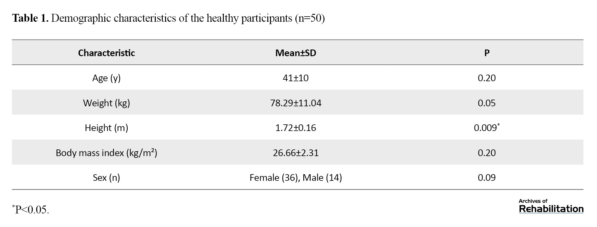
This investigation protocol was granted approval by the Institutional Ethics Committee (IR.USWR.REC.1402.056). Written informed consent was obtained from all participants after they were informed about the study procedures, including the duration of participation and assessment methods. All assessments were conducted by a single physiotherapist, who was aware of the study objectives. Table 1 lists the baseline characteristics of the participants.
Ultrasonography assessment
Ultrasonographic assessment of all cervical muscles was performed using a SIMUT ZinoC4 ultrasound system (Med Fanavaran Plus) equipped with a 4-cm linear transducer. All images were obtained using standardized device settings: probe frequency of 10 MHz, gain of 22, and dynamic range of 75. All other imaging parameters were kept constant throughout the measurements for all participants. To minimize the influence of probe pressure on anterior cervical muscle thickness, an adequate amount of coupling gel and light probe contact were used. Each ultrasonographic measurement was performed three consecutive times. Between measurements, the probe was completely removed from the skin and repositioned at the same anatomical location for subsequent image acquisition. Participants’ positions were maintained throughout the procedure.
Ultrasound evaluation of the cervical multifidus muscle
Considering the importance of cervical lordosis in accurate assessment of neck muscle thickness and to minimize measurement error, the cervical multifidus muscle was examined at the central segment of the cervical lordotic curve, corresponding to the C4 vertebral level [27, 28]. After participant preparation, the spinous landmark corresponding to the C4 vertebral level was identified through manual palpation and subsequently marked. Participants were positioned in a seated posture and instructed to preserve a neutral orientation of the head and cervical region. Due to the anatomical attachment of the cervical multifidus lateral to the spinous processes, the ultrasound probe was initially placed transversely over the spinous processes and then moved laterally. Gentle superior–inferior adjustments were performed until an optimal image of the vertebral lamina and muscle fascia was obtained. At this vertebral level, the cervical multifidus muscle was identified lateral to the spinous landmark, lamina junction and anterior to the semispinalis cervicis muscle.
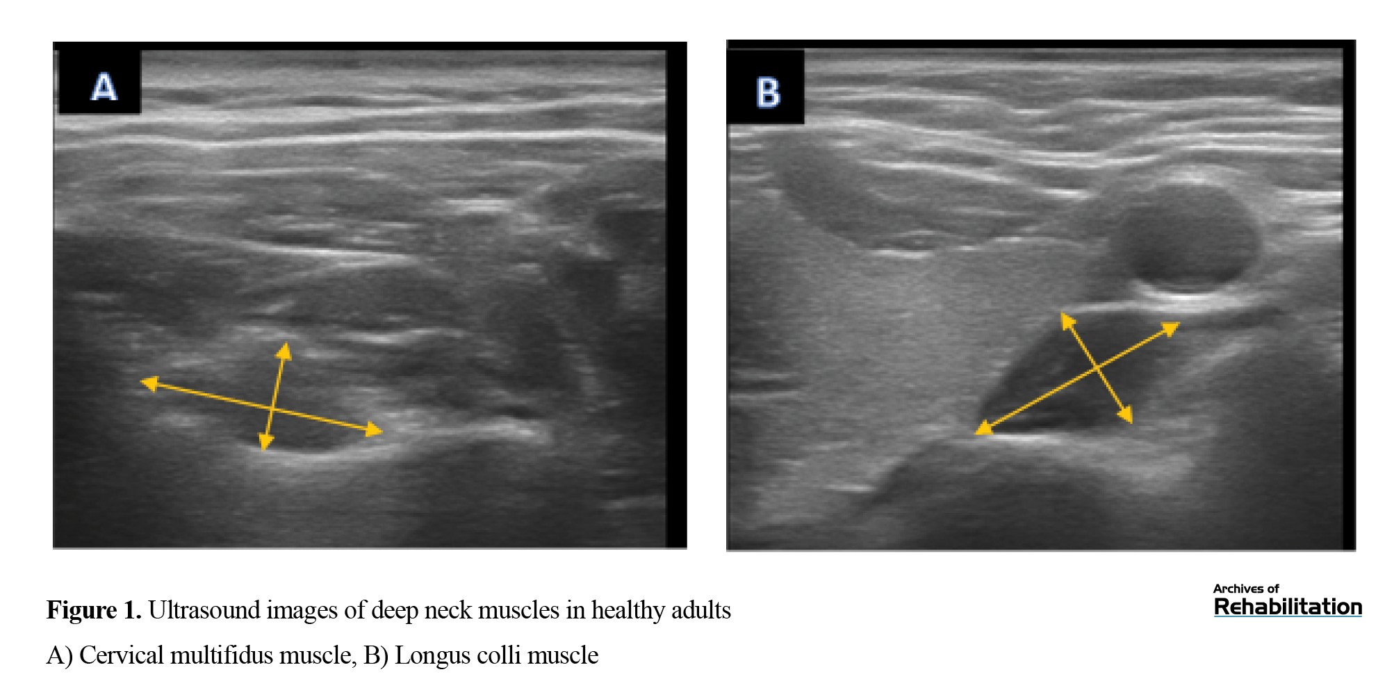
Muscle thickness was measured at rest and operationally defined as the greatest separation between the anterior and posterior fascial layers [27, 28]. Two dimensions were recorded: the anteroposterior diameter (APD), defined as the greatest span between the anterior and posterior margins of the muscle, and the lateral diameter (LD), defined as the maximal span separating the medial and lateral boundaries of the muscle. Measurements were obtained from the dominant side. To reduce measurement error, the mean derived from three repeated measurements was considered for statistical analysis (Figure 1) [27-29].
Ultrasound evaluation of the longus colli muscle
For assessment targeting of the deep cervical flexor musculature, participants were positioned supine with a small folded towel placed beneath the cervical region to preserve normal cervical lordosis, while the upper extremities were positioned adjacent to the trunk [30]. The longus colli muscle was examined at the C5–C6 spinal segment, as this level provides optimal visualization with minimal superimposition of the longus colli and longus capitis muscles. Anatomically, the cricoid cartilage aligns with the C6 vertebra, while the lower margin of the thyroid cartilage corresponds to the C5 vertebral level [30]. The thyroid cartilage was palpated, and a point 2 cm inferior to it was marked as a reference. The probe was then moved approximately 1 cm laterally to visualize the carotid artery in a transverse orientation, within which the longus colli muscle appeared between the carotid artery and the vertebral bodies.
Muscle thickness was quantified as the maximal distance separating the anterior and posterior fascial layers, while muscle width was determined along a line oriented perpendicular to this measurement (Figure 1) [22, 27, 31]. The average value derived from three consecutive measurements was utilized for data analysis.
Ultrasound evaluation of the SCM muscle
Participants were placed in a supine posture with a folded towel supporting the cervical region, and the head was gently rotated approximately 20–30° to the contralateral side from the side being examine. The probe was moved in a cephalad direction, and transverse images were obtained. Head rotation was applied to improve muscle delineation and access.
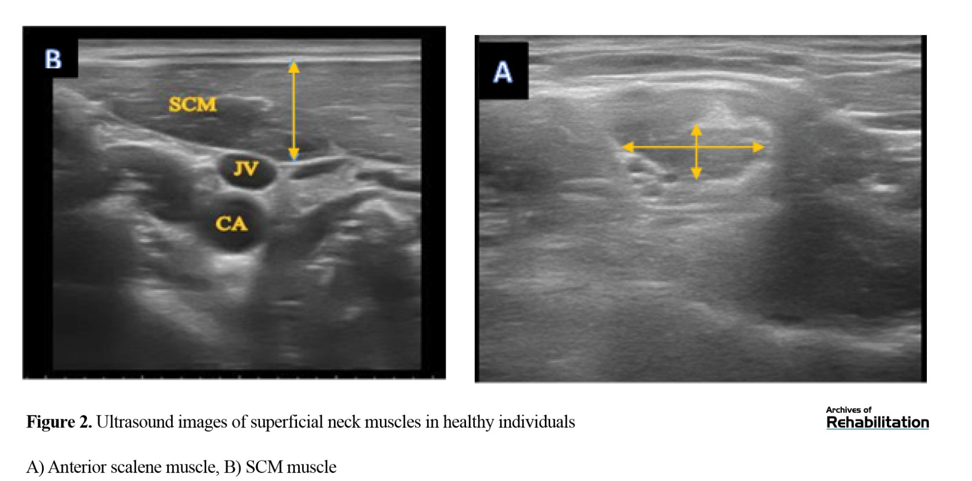
Ultrasound imaging was performed in B-mode using a 10-MHz linear transducer, following the protocol described by Arts et al. [32]. The ultrasound transducer was positioned over the anterolateral aspect of the cervical region, aligned parallel with the midpoint of the muscle, corresponding to the line between the mastoid process and the clavicular attachment. The carotid artery served as an anatomical landmark beneath the sternocleidomastoid muscle, and muscle thickness was quantified as the maximum separation between the anterior and posterior fascial layers (Figure 2).
 The average value obtained from three repeated measurements was used for analysis [32–34].
Ultrasound evaluation of the anterior scalene muscle
For imaging of the scalene muscles, the participant was positioned supine, and the examiner stood on the side being assessed. Participant positioning, examiner position, and probe orientation were identical to those used for SCM assessment. The ultrasound transducer was placed adjacent to the thyroid lobe.
Initially, the brachial plexus roots appeared as oval hypoechoic structures positioned within the interval separating the anterior and middle scalene muscles. The probe was then moved slightly superiorly to visualize the interscalene portion of the brachial plexus at the C6 level, allowing clear identification of the anterior scalene muscle. After each measurement, the probe was removed from the skin. A sufficient amount of gel and minimal probe pressure were applied to minimize tissue compression (Figure 2). The mean value derived from three repeated measurements was considered for statistical evaluation. [30, 35-38].
Spirometry procedure
Respiratory parameters were assessed using a spirometer (Bionet, SPM300, Korea). Spirometry was conducted in compliance with guidelines issued by the American Thoracic Society and the European Respiratory Society (ATS/ERS) [39]. The evaluated indices comprised FVC, forced expiratory volume in 1 second (FEV₁), and the FEV₁/FVC ratio. All measurements were conducted while participants were seated, maintaining an upright trunk posture with the feet resting flat on the floor, the upper limbs relaxed alongside the body, and the head and cervical region aligned in a neutral anatomical position. A chair without a backrest was used to allow unrestricted thoracic movement. Verbal encouragement was provided to ensure maximal effort [40].
To control potential confounding factors, participants were advised to avoid food intake and caffeinated drinks for at least three hours prior to testing and to avoid vigorous physical activity for 24 hours before the test. Smokers and participants with a prior history of tobacco use were excluded. Compliance was verified through direct questioning. Participants were also advised to avoid tight clothing. All spirometry tests were conducted in a clinical hospital environment under stable conditions, and the spirometer was calibrated before data collection [39, 40].
Each participant used a disposable mouthpiece, while a nasal clip was applied to prevent nasal breathing. Participant was asked to take a maximal inhalation followed by a forceful exhalation lasting at least 6 seconds, followed by another deep inspiration [17]. A 5-minute rest period was provided between trials. Testing continued until 3 trials meeting technical acceptability criteria were achieved, and the highest values were recorded for analysis. Trials were repeated if expiration time was insufficient or if inspiratory and expiratory maneuvers were not performed consecutively [41]. Acceptable trials were defined as those with the highest FVC and FEV₁ values, provided that the difference between consecutive trials did not exceed 150–200 mL [17].
Chest wall expansion measurement
Chest wall mobility was assessed using a flexible measuring tape. Participants stood upright with the feet positioned at shoulder width and the upper limbs resting comfortably alongside the body. They were instructed to avoid any additional trunk or limb movements. Measurements were performed with the upper torso uncovered to eliminate restrictions caused by clothing. Chest circumference was measured at two predefined anatomical landmarks, including the anterior axillary line as well as the level of the xiphoid process. The tape was placed horizontally around the chest, with one end stabilized by the examiner. Participants were instructed to execute a maximal deep inhalation followed by a maximal deep exhalation, holding each position for 2 seconds. The numerical difference between thoracic circumference during inspiration and expiration was recorded as chest wall expansion. To enhance measurement reliability, chest wall expansion at each level was measured three consecutive occasions, and the average of the three measurements was used for statistical analysis [19].
Data analysis
All statistical procedures were performed using IBM SPSS version 27.0.1. Descriptive statistics of the study participants’ characteristics, including age, sex, weight, height, and body mass index (BMI), and all primary study variables, were summarized. Indices of central tendency (mean) and variability (standard deviation) were applied to characterize the data. Distributional assumptions were evaluated through application of the Kolmogorov-Smirnov procedure. In addition, to improve the accuracy of normality assessment, skewness and kurtosis indices were calculated, and visual inspection of histograms was conducted. For variables with a normal distribution, associations among variables were examined using the Pearson parametric correlation coefficient. For variables that failed to satisfy normality assumptions, Spearman non‑parametric correlation coefficient was applied.
For statistical reporting, a P value lower 0.05 was defined as statistically significant. Values falling from 0.05 to 0.06, representing a 20% deviation from the conventional significance threshold, was regarded as marginally significant. The magnitude of correlation coefficients was interpreted based on Cohen’s guidelines, in which coefficients of approximately 0.10, 0.30, and 0.50 were interpreted as indicating small, moderate, and large effect sizes, respectively. Following data analysis, all results were reported with two decimal places [42].
Results
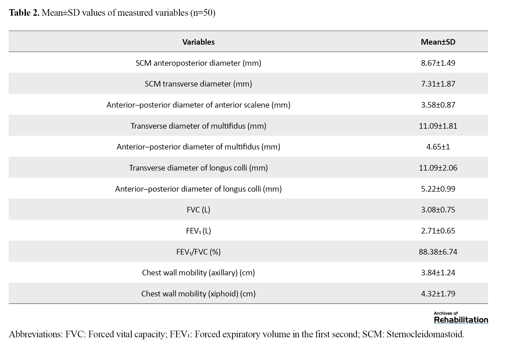
A total of 50 healthy individuals (36 women and 14 men; mean age: 41±10 years) took part in the present investigation. The mean body mass index (BMI) of the participants was 26.66±2.31 kg/m². The distributional normality of demographic variables was assessed through the Kolmogorov-Smirnov test, along with evaluation of skewness, kurtosis, and visual inspection of histograms. The results indicated normal distributions for all demographic variables (Table 1). All primary measured variables demonstrated normal distributions, except for lower chest wall expansion. The average values of the measured variables are presented in Table 2.
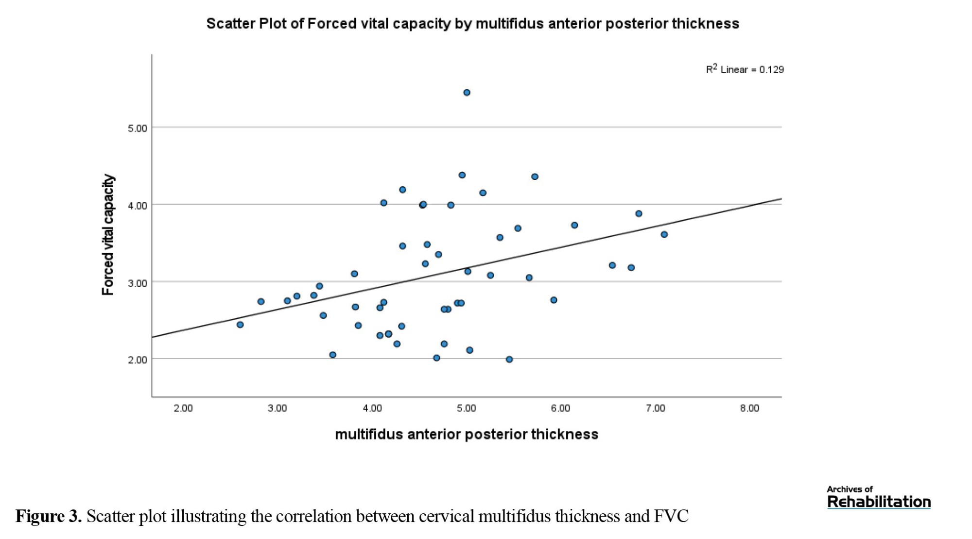
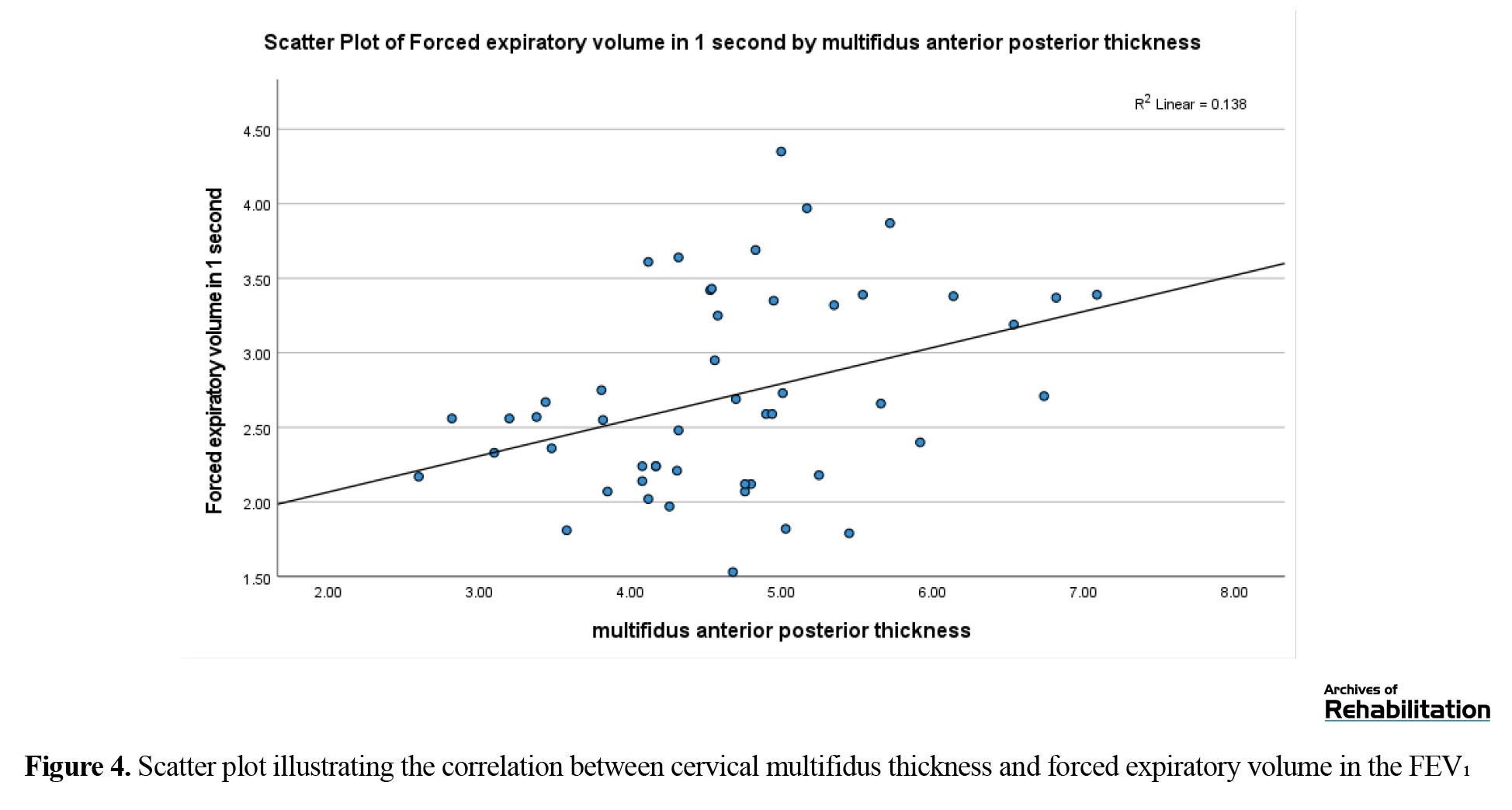
Analysis of the correlation coefficients revealed a statistically significant positive relationship between cervical multifidus muscle thickness and pulmonary function measures, including forced vital capacity (FVC: r=0.35; P=0.01, 95% CI, 0.08%, 0.57% L) as well as forced expiratory volume in one second (FEV₁: r=0.37; P=0.008; 95% CI, 0.01%, 0.59% L), with both associations demonstrating moderate effect magnitudes (Figures 3 and 4).
 No other significant associations were observed between multifidus or longus colli muscle thickness and respiratory volume indices.
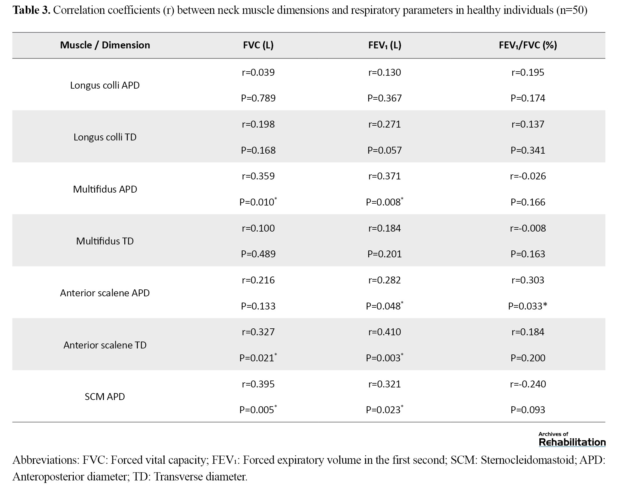
Analysis of the relationships between superficial cervical muscle thickness and spirometric indices showed that the anterior–posterior diameter of the anterior scalene muscle was positively correlated with FEV₁ (r=0.28; P=0.04; 95% CI, 0.001%, 0.52% L) and the FEV₁/FVC ratio (r=0.30; P=0.03; 95% CI, 0.02%, 0.53% L). In addition, the transverse diameter of the anterior scalene muscle demonstrated significant positive correlations with FVC (r= 0.32; P=0.02; 95% CI, 0.04%, 0.55% L) and FEV₁ (r=0.41; P=0.03; 95% CI: 0.15%, 0.62% L), both indicating moderate effect sizes. Furthermore, SCM muscle thickness showed significant positive associations with FVC (r=0.39; P=0.005; 95% CI, 0.13%, 0.6% L) and FEV₁ (r=0.32; P=0.002; 95% CI, 0.04%, 0.55% L) (Table 3).
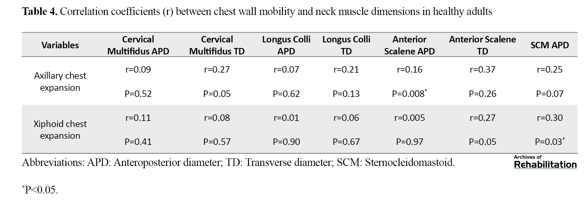
Regarding chest wall mobility, the results demonstrated significant positive correlations between lower chest wall expansion and anterior scalene muscle thickness (r=0.16; P=0.008; 95% CI, 0.12%, 0.42%), as well as SCM muscle thickness (r=0.30; P=0.03; 95% CI, 0.02%, 0.53%) in healthy individuals (Table 4).
Given the relatively moderate sample size (n=50), the study may have lacked adequate statistical power to detect weak correlations. Accordingly, non‑significant results should be viewed cautiously, and further investigations involving larger samples are advised.
Discussion
The principal outcomes of the current investigation indicated that, in healthy individuals, the thickness of certain cervical muscles—specifically the SCM, anterior scalene, and cervical multifidus—was positively associated with spirometric indices. In addition, superficial cervical muscles were related to chest wall mobility. In contrast, no statistically meaningful relationships were observed between longus colli muscle thickness and respiratory parameters. These findings suggest that even in a pain-free and asymptomatic population, structural characteristics of cervical muscles may be linked to respiratory function.
The spirometric values obtained in the present study were within the normal ranges previously reported for healthy individuals. In populations without respiratory dysfunction, the FEV₁/FVC ratio is typically greater than 80%, reflecting normal airway function [40, 43]. Consistent with this, the mean FVC and FEV₁ values in the current study were comparable to reference values described by Quanjer et al. [44] and Stanojevic et al. [45], confirming the adequacy of participant selection and the absence of pulmonary impairment. Therefore, the spirometric data obtained may serve as valid reference values for healthy adults and provide a meaningful basis for comparison with clinical populations or individuals with musculoskeletal disorders.
Similarly, ultrasonographic measurements of cervical muscles, including the multifidus, longus colli, SCM, and anterior scalene, were consistent with values previously reported in healthy populations [21, 32, 46]. Intrinsic cervical muscles, including the multifidus and longus colli, primarily contribute to segmental stability and postural control, whereas the SCM and scalene muscles are more superficial and are involved in dynamic cervical movements and accessory inspiration [3, 21]. These findings support the validity of the ultrasonographic measurements and suggest that the obtained values may be used as baseline data in future investigations.
A key finding of this study was the significant positive association between cervical muscle thickness (SCM, anterior scalene, and multifidus) and spirometric indices, including FVC, FEV₁, and the FEV₁/FVC ratio. Furthermore, SCM and anterior scalene thickness were positively correlated with chest wall mobility at both upper and lower thoracic levels during deep breathing. These relationships may be explained through several anatomical and biomechanical mechanisms. From an anatomical perspective, the scalene and SCM muscles function as accessory inspiratory muscles and directly contribute to thoracic expansion. The scalene muscles originate from the cervical spinal segments and attach to the first and second ribs, facilitating rib elevation during inspiration. Electromyographic studies have demonstrated increased activation of these muscles under conditions of elevated respiratory demand, indicating their role in augmenting ventilation [47]. Consequently, variations in their thickness or function may influence lung volumes such as FVC and FEV₁.
Similarly, the SCM muscle, extending from the mastoid process to the sternum and clavicle, elevates the shoulder girdle during contraction, thereby increasing thoracic volume during inspiration. Increased SCM thickness may therefore reflect greater participation of this muscle in the inspiratory process and can be interpreted within its established role as an accessory respiratory muscle [2, 47]. In addition, the cervical multifidus plays an essential function in maintaining cervical spine stability and facilitating coordinated movement between the neck and thoracic cage. Optimal cervical and trunk alignment may promote effective rib motion and diaphragmatic function. Recent evidence suggests that deep cervical muscle performance and postural control can influence respiratory mechanics [48]. which may explain the observed association between multifidus thickness and spirometric indices.
From a fascial perspective, the cervical region, thoracic cage, and diaphragm are interconnected through a continuous fascial network. This mechanical continuity may facilitate force transmission and coordinated movement between cervical muscles and thoracic structures [2, 49], Such fascial interconnections may represent an additional mechanism underlying the observed associations between cervical muscle thickness and chest wall mobility.
In the context of the present investigation, cervical multifidus thickness demonstrated an association with spirometric indices (FVC and FEV₁), whereas no significant relationship was observed with chest wall mobility. This finding is consistent with the functional role of the multifidus as a deep stabilizing muscle primarily involved in postural control and segmental stability rather than direct thoracic motion [5, 50]. Adequate stability of the cervical spine and upper trunk may enhance respiratory efficiency and intrathoracic pressure generation [51], which is more likely to be reflected in airflow- and volume-related indices than in chest wall excursion.
In contrast, the longus colli muscle does not demonstrate significant associations with spirometric parameters or chest wall mobility. This finding may be explained by its specialized function as a deep cervical flexor and anterior stabilizer [5]. The longus colli primarily contributes to fine motor control, segmental stability, and maintenance of cervical alignment during static tasks, with minimal involvement in ventilatory mechanics or thoracic movement [13]. Unlike superficial cervical muscles that are recruited during increased respiratory demand [47], longus colli activity is predominantly postural in nature, and morphological variations in this muscle are therefore unlikely to be reflected in respiratory indices.
From a neurophysiological standpoint, strong interactions exist between cervical motor control and respiratory function. Motor neurons arising from the lower cervical spinal cord, particularly at the C3-C5 levels, form the phrenic nerve, which innervates the diaphragm. Previous studies have demonstrated that mechanical stimulation or nociceptive input from the cervical region can alter breathing patterns through shared sensorimotor pathways [52, 53]. In addition, proprioceptive input from deep cervical muscles contributes to postural regulation and respiratory coordination, such that impaired cervical stability may influence respiratory performance. However, the direction and causality of these relationships remain to be clarified through longitudinal or interventional studies [25].
Overall, the present findings suggest that cervical muscles are not solely involved in head posture and neck movement but are part of a broader musculoskeletal–neural network contributing to respiratory function. This anatomical and physiological integration may explain the observed positive associations between cervical muscle thickness, spirometric indices, as well as chest wall mobility among healthy individuals.
In conclusion, the findings of the current investigation are aligned with the predefined study objectives. Certain cervical muscles—particularly accessory inspiratory muscles—demonstrate significant associations with spirometric indices and chest wall mobility, whereas such relationships are not observed for all cervical muscles. These differences highlight the distinct functional roles played by superficial versus deep cervical muscles in relation to respiratory function.
Conclusion
The present findings indicate that the thickness of specific cervical muscles, including the SCM, anterior scalene, and multifidus, is positively associated with spirometric indices as well as chest wall mobility among healthy individuals. These findings indicate a potential structural–functional relationship between cervical muscle characteristics and respiratory function. Cervical muscles, particularly the accessory inspiratory muscles, may contribute to respiratory performance through coordinated interactions with the thoracic cage. However, given the cross-sectional nature of this investigation, the results only reflect correlations and do not permit causal inferences or conclusions regarding the effects of interventions. These findings provide baseline data for healthy populations and may guide the design of future longitudinal or interventional studies to explore the clinical relevance of cervical muscles in respiratory function. The study specifically addressed the research questions concerning the relationship between cervical muscle characteristics, spirometric indices, and chest wall mobility in healthy adults.
Study limitations
This investigation has several constraints, as its cross‑sectional design framework limits the capacity to determine cause‑and‑effect relations among cervical muscle characteristics and respiratory function. The study population consisted solely of healthy individuals, so generalization to clinical populations should be made with caution. Convenience sampling was used, which may introduce selection bias. All ultrasonographic measurements were conducted by a single examiner in a single session, so measurement error and dependence on examiner skill (intra-rater reliability) might have influenced the findings. The absence of repeated measurements prevented reporting reliability indices. Future research employing longitudinal approaches, expanded sample sizes, and assessment of measurement reliability are recommended. Investigating clinical populations with cervical musculoskeletal disorders or respiratory conditions would also help generalize the findings and provide normative clinical data.
Ethical Considerations
Compliance with ethical guidelines
This study was approved by the Ethics Committee of the University of Social Welfare and Rehabilitation Sciences, Tehran, Iran (Code: IR.USWR.REC.1402.056). Written informed consent was obtained from all participants before the study, and they were fully informed about the study objectives and procedures. Ethical principles, including confidentiality, voluntary participation with the right to withdraw at any time, no financial burden, and provision for compensating potential harms, were strictly adhered to.
Funding
This article funded by Department of Physiotherapy, University of Social Welfare and Rehabilitation Sciences, Tehran, Iran.
Authors' contributions
Conceptualization: Sima Hatamvand, Nahid Rahmani, and Mohammad Ali Mohseni Bandpei; Methodology: Sima Hatamvand, Nahid Rahmani, and Mohammad Ali Mohseni Bandpei; Validation: Sima Hatamvand and Nahid Rahmani; Formal analysis: Mohammad Saatchi and Sima Hatamvand; Investigation: Sima Hatamvand; Writing, review, and editing: Nahid Rahmani, Mohammad Ali Mohseni Bandpei, Sima Hatamvand, Mohammad Saatchi, and Somayeh Amiri Arimi; Visualization: Sima Hatamvand; Supervision: Nahid Rahmani and Mohammad Ali Mohseni Bandpei; Project administration: Nahid Rahmani and Mohammad Ali Mohseni Bandpei.
Conflict of interest
The authors declared no conflict of interest.
Acknowledgments
The authors would like to express their sincere gratitude to Shahid Rahimi Hospital, Lorestan, and to all those who supported and contributed to the progress and completion of this research.
References
The cervical region is a key component of the human body, playing an essential role in maintaining postural alignment, head balance, and functional mobility [1]. In addition to controlling head and cervical spine movements, cervical muscles also participate in the respiratory process [2, 3]. In particular, superficial structures including the sternocleidomastoid (SCM) and the anterior scalene muscles become active as accessory inspiratory muscles during deep inhalation, contributing to thoracic expansion and facilitating airflow into the lungs [3, 4]. Furthermore, the intrinsic muscle system of the cervical region, with particular emphasis on the longus colli and multifidus, is essential for maintaining cervical spine stability and for ensuring coordinated movement between the neck and trunk [5, 6]. Effective breathing requires adequate cervical spine stability to ensure proper coordination and mechanical efficiency of the muscles involved in respiration [7, 8]. Appropriate stabilization of the cervical region enables respiratory-related muscles, especially accessory inspiratory muscles, to effectively contribute to rib motion and chest wall expansion [5, 9]. Under such conditions, sufficient integrity of the cervical and thoracic spinal segments becomes necessary for maintaining postural control and motor coordination [10, 11]. This stability is largely achieved through the coordinated function of deep cervical and trunk muscles [12, 13]. In light of the involvement of deep cervical muscles in segmental stabilization [13], evaluating the characteristics of these muscles alongside respiratory indices may provide deeper insight into the interaction between the musculoskeletal and respiratory systems.
Growing scientific interest has recently focused on the association between the cervical musculoskeletal system and respiratory function [9, 14]. Evidence suggests that in certain musculoskeletal disorders, such as chronic neck pain, alterations in cervical muscle activation patterns and strength may be associated with reduced chest wall mobility and impaired pulmonary ventilation [15, 16]. Although spirometry provides valuable information regarding airflow and lung volumes [17], chest wall mobility reflects the mechanical function of the respiratory system and can demonstrate structural characteristics and volumetric changes of the thoracic wall during inspiration and expiration [18, 19]. Given the critical role of chest wall motion in effective pulmonary ventilation [20], examining the association between cervical muscle characteristics and chest wall mobility may enhance understanding of the interaction between the respiratory and movement systems.
Ultrasonography represents a non-invasive and accurate imaging technique that allows precise assessment of muscle thickness and morphology [21, 22]. In parallel, spirometry serves as the gold-standard approach for the assessment of respiratory capacity, as reflected by parameters such as forced vital capacity (FVC), forced expiratory volume in one second (FEV₁), and the FEV₁‑to‑FVC ratio regarded as central measures of ventilatory performance [17]. In addition to these tools, assessing chest wall mobility may provide a comprehensive perspective on the mechanical performance of the respiratory system [15].
Despite growing interest in the relationship between the cervical musculoskeletal system and respiratory function, based on comprehensive literature search [23, 24], no study has ever specifically examined the simultaneous association between cervical muscle characteristics (including muscle thickness), spirometric indices, together with chest wall mobility across both asymptomatic individuals and populations with musculoskeletal pain. Although several previous studies have investigated patients with neck pain or other musculoskeletal disorders [23, 25], their focus has primarily been on respiratory or muscular variables in isolation, without integrating assessments of cervical muscle thickness, pulmonary function, and chest wall mobility. Consequently, a research gap exists in this field. Establishing normative data in this area could enhance understanding of the contribution of cervical muscles in respiration and support the development of effective rehabilitation interventions. Therefore, this investigation aims to investigate the association between both intrinsic and superficial cervical muscle thickness, spirometric indices, and chest wall mobility in healthy individuals. It is anticipated that the results generated herein will help clarify the underlying interplay between cervical muscle function and the respiratory system and provide a foundation for future research in clinical populations.
Materials and Methods
Using a cross-sectional design, this investigation recruited 50 asymptomatic individuals aged 20 to 55 years. Sample size estimation was performed through the application of use of Pearson correlation parameters applying Fisher z‑based approach, with reference to the study by Colak et al. [24]. Assuming a correlation coefficient of 0.465, an α threshold of 0.05 and a target statistical power of 80%, a minimum sample size of 34 participants was estimated. To improve the precision of the study, the final study cohort comprised 50 individuals. The selected age range was intended to minimize the influence of skeletal growth-related changes as well as age-related degenerative alterations on cervical muscle structure and respiratory function. The study was conducted between August 2024 and June 2025 in a hospital clinical setting. Participants were recruited through non-probability convenience sampling on a voluntary basis using posters and announcements displayed at the University of Medical Sciences and affiliated public centers. Eligible individuals were enrolled consecutively after screening according to prospectively established eligibility requirements, which included the absence of pain in the spinal column or thoracic region and no history of respiratory diseases. The exclusion criteria included being an athlete (defined as individuals with a regular exercise program), smoking, history of acute trauma, fractures, or surgical procedures involving the spine, thorax, or limbs, presence of any musculoskeletal deformities, forward head posture, or evident scoliosis of the spine or limbs based on clinical assessment by a physiotherapist, cardiovascular, pulmonary, rheumatologic, systemic, or metabolic diseases, malignancies or infections, neurological disorders or neuropathies, pregnancy, psychiatric disorders based on medical history or reported medication use, and a body mass index (BMI) < 20 or > 30 kg/m² [14, 26].
This investigation protocol was granted approval by the Institutional Ethics Committee (IR.USWR.REC.1402.056). Written informed consent was obtained from all participants after they were informed about the study procedures, including the duration of participation and assessment methods. All assessments were conducted by a single physiotherapist, who was aware of the study objectives. Table 1 lists the baseline characteristics of the participants.
Ultrasonography assessment
Ultrasonographic assessment of all cervical muscles was performed using a SIMUT ZinoC4 ultrasound system (Med Fanavaran Plus) equipped with a 4-cm linear transducer. All images were obtained using standardized device settings: probe frequency of 10 MHz, gain of 22, and dynamic range of 75. All other imaging parameters were kept constant throughout the measurements for all participants. To minimize the influence of probe pressure on anterior cervical muscle thickness, an adequate amount of coupling gel and light probe contact were used. Each ultrasonographic measurement was performed three consecutive times. Between measurements, the probe was completely removed from the skin and repositioned at the same anatomical location for subsequent image acquisition. Participants’ positions were maintained throughout the procedure.
Ultrasound evaluation of the cervical multifidus muscle
Considering the importance of cervical lordosis in accurate assessment of neck muscle thickness and to minimize measurement error, the cervical multifidus muscle was examined at the central segment of the cervical lordotic curve, corresponding to the C4 vertebral level [27, 28]. After participant preparation, the spinous landmark corresponding to the C4 vertebral level was identified through manual palpation and subsequently marked. Participants were positioned in a seated posture and instructed to preserve a neutral orientation of the head and cervical region. Due to the anatomical attachment of the cervical multifidus lateral to the spinous processes, the ultrasound probe was initially placed transversely over the spinous processes and then moved laterally. Gentle superior–inferior adjustments were performed until an optimal image of the vertebral lamina and muscle fascia was obtained. At this vertebral level, the cervical multifidus muscle was identified lateral to the spinous landmark, lamina junction and anterior to the semispinalis cervicis muscle.
Muscle thickness was measured at rest and operationally defined as the greatest separation between the anterior and posterior fascial layers [27, 28]. Two dimensions were recorded: the anteroposterior diameter (APD), defined as the greatest span between the anterior and posterior margins of the muscle, and the lateral diameter (LD), defined as the maximal span separating the medial and lateral boundaries of the muscle. Measurements were obtained from the dominant side. To reduce measurement error, the mean derived from three repeated measurements was considered for statistical analysis (Figure 1) [27-29].
Ultrasound evaluation of the longus colli muscle
For assessment targeting of the deep cervical flexor musculature, participants were positioned supine with a small folded towel placed beneath the cervical region to preserve normal cervical lordosis, while the upper extremities were positioned adjacent to the trunk [30]. The longus colli muscle was examined at the C5–C6 spinal segment, as this level provides optimal visualization with minimal superimposition of the longus colli and longus capitis muscles. Anatomically, the cricoid cartilage aligns with the C6 vertebra, while the lower margin of the thyroid cartilage corresponds to the C5 vertebral level [30]. The thyroid cartilage was palpated, and a point 2 cm inferior to it was marked as a reference. The probe was then moved approximately 1 cm laterally to visualize the carotid artery in a transverse orientation, within which the longus colli muscle appeared between the carotid artery and the vertebral bodies.
Muscle thickness was quantified as the maximal distance separating the anterior and posterior fascial layers, while muscle width was determined along a line oriented perpendicular to this measurement (Figure 1) [22, 27, 31]. The average value derived from three consecutive measurements was utilized for data analysis.
Ultrasound evaluation of the SCM muscle
Participants were placed in a supine posture with a folded towel supporting the cervical region, and the head was gently rotated approximately 20–30° to the contralateral side from the side being examine. The probe was moved in a cephalad direction, and transverse images were obtained. Head rotation was applied to improve muscle delineation and access.
Ultrasound imaging was performed in B-mode using a 10-MHz linear transducer, following the protocol described by Arts et al. [32]. The ultrasound transducer was positioned over the anterolateral aspect of the cervical region, aligned parallel with the midpoint of the muscle, corresponding to the line between the mastoid process and the clavicular attachment. The carotid artery served as an anatomical landmark beneath the sternocleidomastoid muscle, and muscle thickness was quantified as the maximum separation between the anterior and posterior fascial layers (Figure 2).
Ultrasound evaluation of the anterior scalene muscle
For imaging of the scalene muscles, the participant was positioned supine, and the examiner stood on the side being assessed. Participant positioning, examiner position, and probe orientation were identical to those used for SCM assessment. The ultrasound transducer was placed adjacent to the thyroid lobe.
Initially, the brachial plexus roots appeared as oval hypoechoic structures positioned within the interval separating the anterior and middle scalene muscles. The probe was then moved slightly superiorly to visualize the interscalene portion of the brachial plexus at the C6 level, allowing clear identification of the anterior scalene muscle. After each measurement, the probe was removed from the skin. A sufficient amount of gel and minimal probe pressure were applied to minimize tissue compression (Figure 2). The mean value derived from three repeated measurements was considered for statistical evaluation. [30, 35-38].
Spirometry procedure
Respiratory parameters were assessed using a spirometer (Bionet, SPM300, Korea). Spirometry was conducted in compliance with guidelines issued by the American Thoracic Society and the European Respiratory Society (ATS/ERS) [39]. The evaluated indices comprised FVC, forced expiratory volume in 1 second (FEV₁), and the FEV₁/FVC ratio. All measurements were conducted while participants were seated, maintaining an upright trunk posture with the feet resting flat on the floor, the upper limbs relaxed alongside the body, and the head and cervical region aligned in a neutral anatomical position. A chair without a backrest was used to allow unrestricted thoracic movement. Verbal encouragement was provided to ensure maximal effort [40].
To control potential confounding factors, participants were advised to avoid food intake and caffeinated drinks for at least three hours prior to testing and to avoid vigorous physical activity for 24 hours before the test. Smokers and participants with a prior history of tobacco use were excluded. Compliance was verified through direct questioning. Participants were also advised to avoid tight clothing. All spirometry tests were conducted in a clinical hospital environment under stable conditions, and the spirometer was calibrated before data collection [39, 40].
Each participant used a disposable mouthpiece, while a nasal clip was applied to prevent nasal breathing. Participant was asked to take a maximal inhalation followed by a forceful exhalation lasting at least 6 seconds, followed by another deep inspiration [17]. A 5-minute rest period was provided between trials. Testing continued until 3 trials meeting technical acceptability criteria were achieved, and the highest values were recorded for analysis. Trials were repeated if expiration time was insufficient or if inspiratory and expiratory maneuvers were not performed consecutively [41]. Acceptable trials were defined as those with the highest FVC and FEV₁ values, provided that the difference between consecutive trials did not exceed 150–200 mL [17].
Chest wall expansion measurement
Chest wall mobility was assessed using a flexible measuring tape. Participants stood upright with the feet positioned at shoulder width and the upper limbs resting comfortably alongside the body. They were instructed to avoid any additional trunk or limb movements. Measurements were performed with the upper torso uncovered to eliminate restrictions caused by clothing. Chest circumference was measured at two predefined anatomical landmarks, including the anterior axillary line as well as the level of the xiphoid process. The tape was placed horizontally around the chest, with one end stabilized by the examiner. Participants were instructed to execute a maximal deep inhalation followed by a maximal deep exhalation, holding each position for 2 seconds. The numerical difference between thoracic circumference during inspiration and expiration was recorded as chest wall expansion. To enhance measurement reliability, chest wall expansion at each level was measured three consecutive occasions, and the average of the three measurements was used for statistical analysis [19].
Data analysis
All statistical procedures were performed using IBM SPSS version 27.0.1. Descriptive statistics of the study participants’ characteristics, including age, sex, weight, height, and body mass index (BMI), and all primary study variables, were summarized. Indices of central tendency (mean) and variability (standard deviation) were applied to characterize the data. Distributional assumptions were evaluated through application of the Kolmogorov-Smirnov procedure. In addition, to improve the accuracy of normality assessment, skewness and kurtosis indices were calculated, and visual inspection of histograms was conducted. For variables with a normal distribution, associations among variables were examined using the Pearson parametric correlation coefficient. For variables that failed to satisfy normality assumptions, Spearman non‑parametric correlation coefficient was applied.
For statistical reporting, a P value lower 0.05 was defined as statistically significant. Values falling from 0.05 to 0.06, representing a 20% deviation from the conventional significance threshold, was regarded as marginally significant. The magnitude of correlation coefficients was interpreted based on Cohen’s guidelines, in which coefficients of approximately 0.10, 0.30, and 0.50 were interpreted as indicating small, moderate, and large effect sizes, respectively. Following data analysis, all results were reported with two decimal places [42].
Results
A total of 50 healthy individuals (36 women and 14 men; mean age: 41±10 years) took part in the present investigation. The mean body mass index (BMI) of the participants was 26.66±2.31 kg/m². The distributional normality of demographic variables was assessed through the Kolmogorov-Smirnov test, along with evaluation of skewness, kurtosis, and visual inspection of histograms. The results indicated normal distributions for all demographic variables (Table 1). All primary measured variables demonstrated normal distributions, except for lower chest wall expansion. The average values of the measured variables are presented in Table 2.
Analysis of the correlation coefficients revealed a statistically significant positive relationship between cervical multifidus muscle thickness and pulmonary function measures, including forced vital capacity (FVC: r=0.35; P=0.01, 95% CI, 0.08%, 0.57% L) as well as forced expiratory volume in one second (FEV₁: r=0.37; P=0.008; 95% CI, 0.01%, 0.59% L), with both associations demonstrating moderate effect magnitudes (Figures 3 and 4).
Analysis of the relationships between superficial cervical muscle thickness and spirometric indices showed that the anterior–posterior diameter of the anterior scalene muscle was positively correlated with FEV₁ (r=0.28; P=0.04; 95% CI, 0.001%, 0.52% L) and the FEV₁/FVC ratio (r=0.30; P=0.03; 95% CI, 0.02%, 0.53% L). In addition, the transverse diameter of the anterior scalene muscle demonstrated significant positive correlations with FVC (r= 0.32; P=0.02; 95% CI, 0.04%, 0.55% L) and FEV₁ (r=0.41; P=0.03; 95% CI: 0.15%, 0.62% L), both indicating moderate effect sizes. Furthermore, SCM muscle thickness showed significant positive associations with FVC (r=0.39; P=0.005; 95% CI, 0.13%, 0.6% L) and FEV₁ (r=0.32; P=0.002; 95% CI, 0.04%, 0.55% L) (Table 3).
Regarding chest wall mobility, the results demonstrated significant positive correlations between lower chest wall expansion and anterior scalene muscle thickness (r=0.16; P=0.008; 95% CI, 0.12%, 0.42%), as well as SCM muscle thickness (r=0.30; P=0.03; 95% CI, 0.02%, 0.53%) in healthy individuals (Table 4).
Given the relatively moderate sample size (n=50), the study may have lacked adequate statistical power to detect weak correlations. Accordingly, non‑significant results should be viewed cautiously, and further investigations involving larger samples are advised.
Discussion
The principal outcomes of the current investigation indicated that, in healthy individuals, the thickness of certain cervical muscles—specifically the SCM, anterior scalene, and cervical multifidus—was positively associated with spirometric indices. In addition, superficial cervical muscles were related to chest wall mobility. In contrast, no statistically meaningful relationships were observed between longus colli muscle thickness and respiratory parameters. These findings suggest that even in a pain-free and asymptomatic population, structural characteristics of cervical muscles may be linked to respiratory function.
The spirometric values obtained in the present study were within the normal ranges previously reported for healthy individuals. In populations without respiratory dysfunction, the FEV₁/FVC ratio is typically greater than 80%, reflecting normal airway function [40, 43]. Consistent with this, the mean FVC and FEV₁ values in the current study were comparable to reference values described by Quanjer et al. [44] and Stanojevic et al. [45], confirming the adequacy of participant selection and the absence of pulmonary impairment. Therefore, the spirometric data obtained may serve as valid reference values for healthy adults and provide a meaningful basis for comparison with clinical populations or individuals with musculoskeletal disorders.
Similarly, ultrasonographic measurements of cervical muscles, including the multifidus, longus colli, SCM, and anterior scalene, were consistent with values previously reported in healthy populations [21, 32, 46]. Intrinsic cervical muscles, including the multifidus and longus colli, primarily contribute to segmental stability and postural control, whereas the SCM and scalene muscles are more superficial and are involved in dynamic cervical movements and accessory inspiration [3, 21]. These findings support the validity of the ultrasonographic measurements and suggest that the obtained values may be used as baseline data in future investigations.
A key finding of this study was the significant positive association between cervical muscle thickness (SCM, anterior scalene, and multifidus) and spirometric indices, including FVC, FEV₁, and the FEV₁/FVC ratio. Furthermore, SCM and anterior scalene thickness were positively correlated with chest wall mobility at both upper and lower thoracic levels during deep breathing. These relationships may be explained through several anatomical and biomechanical mechanisms. From an anatomical perspective, the scalene and SCM muscles function as accessory inspiratory muscles and directly contribute to thoracic expansion. The scalene muscles originate from the cervical spinal segments and attach to the first and second ribs, facilitating rib elevation during inspiration. Electromyographic studies have demonstrated increased activation of these muscles under conditions of elevated respiratory demand, indicating their role in augmenting ventilation [47]. Consequently, variations in their thickness or function may influence lung volumes such as FVC and FEV₁.
Similarly, the SCM muscle, extending from the mastoid process to the sternum and clavicle, elevates the shoulder girdle during contraction, thereby increasing thoracic volume during inspiration. Increased SCM thickness may therefore reflect greater participation of this muscle in the inspiratory process and can be interpreted within its established role as an accessory respiratory muscle [2, 47]. In addition, the cervical multifidus plays an essential function in maintaining cervical spine stability and facilitating coordinated movement between the neck and thoracic cage. Optimal cervical and trunk alignment may promote effective rib motion and diaphragmatic function. Recent evidence suggests that deep cervical muscle performance and postural control can influence respiratory mechanics [48]. which may explain the observed association between multifidus thickness and spirometric indices.
From a fascial perspective, the cervical region, thoracic cage, and diaphragm are interconnected through a continuous fascial network. This mechanical continuity may facilitate force transmission and coordinated movement between cervical muscles and thoracic structures [2, 49], Such fascial interconnections may represent an additional mechanism underlying the observed associations between cervical muscle thickness and chest wall mobility.
In the context of the present investigation, cervical multifidus thickness demonstrated an association with spirometric indices (FVC and FEV₁), whereas no significant relationship was observed with chest wall mobility. This finding is consistent with the functional role of the multifidus as a deep stabilizing muscle primarily involved in postural control and segmental stability rather than direct thoracic motion [5, 50]. Adequate stability of the cervical spine and upper trunk may enhance respiratory efficiency and intrathoracic pressure generation [51], which is more likely to be reflected in airflow- and volume-related indices than in chest wall excursion.
In contrast, the longus colli muscle does not demonstrate significant associations with spirometric parameters or chest wall mobility. This finding may be explained by its specialized function as a deep cervical flexor and anterior stabilizer [5]. The longus colli primarily contributes to fine motor control, segmental stability, and maintenance of cervical alignment during static tasks, with minimal involvement in ventilatory mechanics or thoracic movement [13]. Unlike superficial cervical muscles that are recruited during increased respiratory demand [47], longus colli activity is predominantly postural in nature, and morphological variations in this muscle are therefore unlikely to be reflected in respiratory indices.
From a neurophysiological standpoint, strong interactions exist between cervical motor control and respiratory function. Motor neurons arising from the lower cervical spinal cord, particularly at the C3-C5 levels, form the phrenic nerve, which innervates the diaphragm. Previous studies have demonstrated that mechanical stimulation or nociceptive input from the cervical region can alter breathing patterns through shared sensorimotor pathways [52, 53]. In addition, proprioceptive input from deep cervical muscles contributes to postural regulation and respiratory coordination, such that impaired cervical stability may influence respiratory performance. However, the direction and causality of these relationships remain to be clarified through longitudinal or interventional studies [25].
Overall, the present findings suggest that cervical muscles are not solely involved in head posture and neck movement but are part of a broader musculoskeletal–neural network contributing to respiratory function. This anatomical and physiological integration may explain the observed positive associations between cervical muscle thickness, spirometric indices, as well as chest wall mobility among healthy individuals.
In conclusion, the findings of the current investigation are aligned with the predefined study objectives. Certain cervical muscles—particularly accessory inspiratory muscles—demonstrate significant associations with spirometric indices and chest wall mobility, whereas such relationships are not observed for all cervical muscles. These differences highlight the distinct functional roles played by superficial versus deep cervical muscles in relation to respiratory function.
Conclusion
The present findings indicate that the thickness of specific cervical muscles, including the SCM, anterior scalene, and multifidus, is positively associated with spirometric indices as well as chest wall mobility among healthy individuals. These findings indicate a potential structural–functional relationship between cervical muscle characteristics and respiratory function. Cervical muscles, particularly the accessory inspiratory muscles, may contribute to respiratory performance through coordinated interactions with the thoracic cage. However, given the cross-sectional nature of this investigation, the results only reflect correlations and do not permit causal inferences or conclusions regarding the effects of interventions. These findings provide baseline data for healthy populations and may guide the design of future longitudinal or interventional studies to explore the clinical relevance of cervical muscles in respiratory function. The study specifically addressed the research questions concerning the relationship between cervical muscle characteristics, spirometric indices, and chest wall mobility in healthy adults.
Study limitations
This investigation has several constraints, as its cross‑sectional design framework limits the capacity to determine cause‑and‑effect relations among cervical muscle characteristics and respiratory function. The study population consisted solely of healthy individuals, so generalization to clinical populations should be made with caution. Convenience sampling was used, which may introduce selection bias. All ultrasonographic measurements were conducted by a single examiner in a single session, so measurement error and dependence on examiner skill (intra-rater reliability) might have influenced the findings. The absence of repeated measurements prevented reporting reliability indices. Future research employing longitudinal approaches, expanded sample sizes, and assessment of measurement reliability are recommended. Investigating clinical populations with cervical musculoskeletal disorders or respiratory conditions would also help generalize the findings and provide normative clinical data.
Ethical Considerations
Compliance with ethical guidelines
This study was approved by the Ethics Committee of the University of Social Welfare and Rehabilitation Sciences, Tehran, Iran (Code: IR.USWR.REC.1402.056). Written informed consent was obtained from all participants before the study, and they were fully informed about the study objectives and procedures. Ethical principles, including confidentiality, voluntary participation with the right to withdraw at any time, no financial burden, and provision for compensating potential harms, were strictly adhered to.
Funding
This article funded by Department of Physiotherapy, University of Social Welfare and Rehabilitation Sciences, Tehran, Iran.
Authors' contributions
Conceptualization: Sima Hatamvand, Nahid Rahmani, and Mohammad Ali Mohseni Bandpei; Methodology: Sima Hatamvand, Nahid Rahmani, and Mohammad Ali Mohseni Bandpei; Validation: Sima Hatamvand and Nahid Rahmani; Formal analysis: Mohammad Saatchi and Sima Hatamvand; Investigation: Sima Hatamvand; Writing, review, and editing: Nahid Rahmani, Mohammad Ali Mohseni Bandpei, Sima Hatamvand, Mohammad Saatchi, and Somayeh Amiri Arimi; Visualization: Sima Hatamvand; Supervision: Nahid Rahmani and Mohammad Ali Mohseni Bandpei; Project administration: Nahid Rahmani and Mohammad Ali Mohseni Bandpei.
Conflict of interest
The authors declared no conflict of interest.
Acknowledgments
The authors would like to express their sincere gratitude to Shahid Rahimi Hospital, Lorestan, and to all those who supported and contributed to the progress and completion of this research.
References
- Vos T, Lim SS, Abbafati C, Abbas KM, Abbasi M, Abbasifard M, et al. Global burden of 369 diseases and injuries in 204 countries and territories, 1990-2019: A systematic analysis for the Global Burden of Disease Study 2019. Lancet (London, England). 2020; 396(10258):1204-22 [DOI:10.1016/S0140-6736(20)30925-9] [PMID]
- Bordoni B, Jozsa F, Varacallo MA. Anatomy, Head and Neck: Sternocleidomastoid Muscle. [Updated 2026 Apr 6]. In: StatPearls [Internet]. Treasure Island (FL): StatPearls Publishing; 2026. [Link]
- Falla D, Jull G, Edwards S, Koh K, Rainoldi A. Neuromuscular efficiency of the sternocleidomastoid and anterior scalene muscles in patients with chronic neck pain. Disability and Rehabilitation. 2004; 26(12):712-7. [DOI:10.1080/09638280410001704287] [PMID]
- Falla D, Rainoldi A, Merletti R, Jull G. Myoelectric manifestations of sternocleidomastoid and anterior scalene muscle fatigue in chronic neck pain patients. Clinical Neurophysiology. 2003; 114(3):488-95. [DOI:10.1016/S1388-2457(02)00418-2] [PMID]
- Falla DL, Jull GA, Hodges PW. Patients with neck pain demonstrate reduced electromyographic activity of the deep cervical flexor muscles during performance of the craniocervical flexion test. Spine (Phila Pa 1976). 2004; 29(19):2108-14. [DOI:10.1097/01.brs.0000141170.89317.0e] [PMID]
- Falla D, O’Leary S, Farina D, Jull G. The change in deep cervical flexor activity after training is associated with the degree of pain reduction in patients with chronic neck pain. The Clinical Journal of Pain. 2012; 28(7):628-34 [DOI:10.1097/AJP.0b013e31823e9378] [PMID]
- Kolář P, Šulc J, Kynčl M, Šanda J, Čakrt O, Andel R, et al. Postural function of the diaphragm in persons with and without chronic low back pain. Journal of Orthopaedic & Sports Physical Therapy. 2012; 42(4):352-62. [DOI:10.2519/jospt.2012.3830] [PMID]
- Schmid M, Conforto S, Bibbo D, D’Alessio T. Respiration and postural sway: Detection of phase synchronizations and interactions. Human Movement Science. 2004; 23(2):105-19. [DOI:10.1016/j.humov.2004.06.001] [PMID]
- Kapreli E, Vourazanis E, Billis E, Oldham J, Strimpakos N. Respiratory dysfunction in chronic neck pain patients. A pilot study. Cephalalgia. 2009; 29(7):701-10. [DOI:10.1111/j.1468-2982.2008.01787.x] [PMID]
- Lewit K. Relation of faulty respiration to posture, with clinical implications. The Journal of the American Osteopathic Association. 1980; 79(8):525-9. [PMID]
- Panjabi MM. The stabilizing system of the spine. Part I. Function, dysfunction, adaptation, and enhancement. Journal of Spinal Disorders. 1992; 5(4):383-9. [DOI:10.1097/00002517-199212000-00001] [PMID]
- Jull G, Kristjansson E, Dall’Alba P. Impairment in the cervical flexors: A comparison of whiplash and insidious onset neck pain patients. Manual Therapy. 2004; 9(2):89-94. [DOI:10.1016/S1356-689X(03)00086-9] [PMID]
- Falla D, O’Leary S, Fagan A, Jull G. Recruitment of the deep cervical flexor muscles during a postural-correction exercise performed in sitting. Manual Therapy. 2007; 12(2):139-43. [DOI:10.1016/j.math.2006.06.003] [PMID]
- Dimitriadis Z, Kapreli E, Strimpakos N, Oldham J. Respiratory weakness in patients with chronic neck pain. Manual Therapy. 2013; 18(3):248-53. [DOI:10.1016/j.math.2012.10.014] [PMID]
- Kaneko H, Horie J. Breathing movements of the chest and abdominal wall in healthy subjects. Respiratory Care. 2012; 57(9):1442-51. [DOI:10.4187/respcare.01655] [PMID]
- Cheon JH, Lim NN, Lee GS, Won KH, Lee SH, Kang EY, et al. Differences of spinal curvature, thoracic mobility, and respiratory strength between chronic neck pain patients and people without cervical pain. Annals of Rehabilitation Medicine. 2020; 44(1):58-68. [DOI:10.5535/arm.2020.44.1.58] [PMID]
- Miller MR, Hankinson J, Brusasco V, Burgos F, Casaburi R, Coates A, et al. Standardisation of spirometry. European Respiratory Journal. 2005; 26(2):319-38. [DOI:10.1183/09031936.05.00034805] [PMID]
- Gómez-Recio M, Bastir M, LoMauro A, Aliverti A, Beyer B. Three-dimensional geometric morphometric shape analysis of chest wall kinematics in different breathing conditions. Royal Society Open Science. 2024; 11(7):240548. [DOI:10.1098/rsos.240548] [PMID]
- Malaguti C, Rondelli RR, De Souza LM, Domingues M, Corso SD. Reliability of chest wall mobility and its correlation with pulmonary function in patients with chronic obstructive pulmonary disease. Respiratory Care. 2009; 54(12):1703-11. [DOI:10.4187/respcare.09541703] [PMID]
- Derasse M, Lefebvre S, Liistro G, Reychler G. Chest expansion and lung function for healthy subjects and individuals with pulmonary disease. Respiratory Care. 2021; 66(4):661-8. [DOI:10.4187/respcare.08350] [PMID]
- Javanshir K, Mohseni-Bandpei MA, Rezasoltani A, Amiri M, Rahgozar M. Ultrasonography of longus colli muscle: A reliability study on healthy subjects and patients with chronic neck pain. Journal of Bodywork and Movement Therapies. 2011; 15(1):50-6. [DOI:10.1016/j.jbmt.2009.07.005] [PMID]
- Javanshir K, Rezasoltani A, Mohseni-Bandpei MA, Amiri M, Ortega-Santiago R, Fernández-de-Las-Peñas C. Ultrasound assessment of bilateral longus colli muscles in subjects with chronic bilateral neck pain. American Journal of Physical Medicine & Rehabilitation. 2011; 90(4):293-301. [DOI:10.1097/PHM.0b013e31820173e5] [PMID]
- Hii EY, Kuo YL, Cheng KC, Hung CH, Tsai YJ. Ultrasonographic measurement indicated patients with chronic neck pain had reduced diaphragm thickness and mobility along with declined respiratory functions. Musculoskeletal Science & Practice. 2024; 72:102951. [DOI:10.1016/j.msksp.2024.102951] [PMID]
- Colak GY, Ozyurek S, Sengul YS, Kalemci O. Differences of diaphragmatic muscle contraction between female patients with chronic neck pain and asymptomatic controls: A case-control study based on ultrasonography. Musculoskeletal Science and Practice. 2024; 69:102894. [DOI:10.1016/j.msksp.2023.102894] [PMID]
- Dimitriadis Z, Kapreli E, Strimpakos N, Oldham J. Pulmonary function of patients with chronic neck pain: A spirometry study. Respiratory Care. 2014; 59(4):543-9. [DOI:10.4187/respcare.01828] [PMID]
- Kiryu S, Loring SH, Mori Y, Rofsky NM, Hatabu H, Takahashi M. Quantitative analysis of the velocity and synchronicity of diaphragmatic motion: dynamic MRI in different postures. Magnetic Resonance Imaging. 2006; 24(10):132-32. [DOI:10.1016/j.mri.2006.08.009] [PMID]
- Amiri-Arimi S, Mohseni Bandpei M, Rezasoltani A, Javanshir K, Biglarian A. Measurement of cervical multifidus and longus colli muscle dimensions in patients with cervical radiculopathy and healthy controls using ultrasonography: A reliability study. PM & R. 2019; 11(3):236-42. [DOI:10.1016/j.pmrj.2018.07.014] [PMID]
- Lee JP, Tseng WYI, Shau YW, Wang CL, Wang HK, Wang SF. Measurement of segmental cervical multifidus contraction by ultrasonography in asymptomatic adults. Manual therapy. 2007; 12(3):286-94. [DOI:10.1016/j.math.2006.07.008] [PMID]
- Goodarzi F, Karimi N, Rahnama L, Khodakarim L. Differences in cervical extensor muscles thickness on subjects with normal head posture and forward head posture: an ultrasonography study. Journal of Rehabilitation Sciences & Research. 2015; 2(2):23-6. [DOI:10.30476/jrsr.2015.41069]
- Sidiropoulos G, Strimpakos N, Kanellopoulos AK, Tsekoura M, Alexiou K, Papakonstantinou O, et al. Measurement reliability for the anatomical characteristics of cervical muscles using musculoskeletal ultrasound in healthy individuals. Muscles. 2025; 4(3):28. [DOI:10.3390/muscles4030028] [PMID]
- Zargoosh M, Amiri M, Abdollahi I, Rahnama L, Lak R. [Reliability of longus colli and capitis muscles cross sectional area measurement during cranio cervical flexion test using ultrasonography (Persian)]. Archives of Rehabilitation. 2017; 18(1):43-50. [DOI:10.21859/jrehab-180143]
- Arts IMP, Pillen S, Schelhaas HJ, Overeem S, Zwarts MJ. Normal values for quantitative muscle ultrasonography in adults. Muscle & Nerve. 2010; 41(1):32-41. [DOI:10.1002/mus.21458] [PMID]
- Lee HJ, Song JM. Deep neck flexor and sternocleidomastoid muscle thickness change in persons with no current neck pain using rehabilitative ultrasonograpic imaging. The Journal of Korean Physical Therapy. 2016; 28(6):349-54. [DOI:10.18857/jkpt.2016.28.6.349]
- Shiraishi M, Higashimoto Y, Sugiya R, Mizusawa H, Takeda Y, Fujita S, et al. Sternocleidomastoid muscle thickness correlates with exercise tolerance in patients with COPD. Respiration. 2023; 102(1):64-73. [DOI:10.1159/000527100] [PMID]
- Rached R, Hsing W, Rached C. Evaluation of the efficacy of ropivacaine injection in the anterior and middle scalene muscles guided by ultrasonography in the treatment of Thoracic Outlet Syndrome. Revista da Associacao Medica Brasileira. 2019; 65(7):982-7. [DOI:10.1590/1806-9282.65.7.982] [PMID]
- Bottros MM, AuBuchon JD, McLaughlin LN, Altchek DW, Illig KA, Thompson RW. Exercise-enhanced, ultrasound-guided anterior scalene muscle/pectoralis minor muscle blocks can facilitate the diagnosis of neurogenic thoracic outlet syndrome in the high-performance overhead athlete. The American Journal of Sports Medicine. 2017; 45(1):189-94. [DOI:10.1177/0363546516665801] [PMID]
- Valera-Calero JA, Gómez-Sánchez S, Fernández-de-Las-Peñas C, Plaza-Manzano G, Sanchez-Jorge S, Navarro-Santana MJ. A procedure for measuring anterior scalene morphology and quality with ultrasound imaging: An intra-and inter-rater reliability study. Ultrasound in Medicine and Biology. 2023; 49(8):1817-23. [DOI:10.1016/j.ultrasmedbio.2023.04.005] [PMID]
- Campbell K, Pearl G, Ojukwu O, Grimsley B, Gunn C, Ramamoorthy S. Ultrasonographic changes in the anterior scalene muscle in neurogenic thoracic outlet syndrome. Journal of Vascular Surgery. 2025; 81(5):1131-37. [DOI:10.1016/j.jvs.2025.01.195] [PMID]
- Nicholson PJ. The updated ATS/ERS spirometry technical standards. Occupational Medicine (Oxford, England). 2020; 70(3):146-8. [DOI:10.1093/occmed/kqaa030] [PMID]
- Pellegrino R, Viegi G, Brusasco V, Crapo RO, Burgos F, Casaburi R, et al. Interpretative strategies for lung function tests. The European Respiratory Journal. 2005; 26(5):948-68. [DOI:10.1183/09031936.05.00035205] [PMID]
- Alvisi V, Marangoni E, Zannoli S, Uneddu M, Uggento R, Farabegoli L, et al. Pulmonary function and expiratory flow limitation in acute cervical spinal cord injury. Archives of Physical Medicine and Rehabilitation. 2012; 93(11):1950-6. [DOI:10.1016/j.apmr.2012.04.015] [PMID]
- Benesty J, Chen J, Huang Y, Cohen I. Noise reduction in speech processing. Berlin: Springer Science & Business Media; 2009. [DOI:10.1007/978-3-642-00296-0]
- Rabe KF, Hurd S, Anzueto A, Barnes PJ, Buist SA, Calverley P, et al. Global strategy for the diagnosis, management, and prevention of chronic obstructive pulmonary disease: GOLD executive summary. American Journal of Respiratory and Critical Care Medicine. 2007; 176(6):532-55. [DOI:10.1164/rccm.200703-456so] [PMID]
- Quanjer PH, Stanojevic S, Cole TJ, Baur X, Hall GL, Culver BH, et al. Multi-ethnic reference values for spirometry for the 3-95-yr age range: The global lung function 2012 equations. The European Respiratory Journal. 2012; 40(6):1324-43. [DOI:10.1183/09031936.00080312] [PMID]
- Stanojevic S, Wade A, Stocks J, Hankinson J, Coates AL, Pan H, et al. Reference ranges for spirometry across all ages: A new approach. American Journal of Respiratory and Critical Care Medicine. 2008; 177(3):253-60. [DOI:10.1164/rccm.200708-1248OC] [PMID]
- Valera-Calero JA, Gallego-Sendarrubias GM, Fernández-de-las-Peñas C, Cleland JA, Ortega-Santiago R, Arias-Buría JL. Panoramic ultrasound examination of posterior neck extensors in healthy subjects: Intra-examiner reliability study. Diagnostics (Basel, Switzerland). 2020; 10(10):740. [DOI:10.3390/diagnostics10100740] [PMID]
- Basoudan N, Rodrigues A, Gallina A, Garland J, Guenette JA, Shadgan B, et al. Scalene and sternocleidomastoid activation during normoxic and hypoxic incremental inspiratory loading. Physiological Reports. 2020; 8(14):e14522. [DOI:10.14814/phy2.14522] [PMID]
- Cefalì A, Santini D, Lopez G, Maselli F, Rossettini G, Crestani M, et al. Effects of breathing exercises on neck pain management: A systematic review with meta-analysis. Journal of Clinical Medicine. 2025; 14(3):709. [DOI:10.3390/jcm14030709] [PMID]
- Myers TW. Anatomy trains: Myofascial meridians for manual and movement therapists. 2th ed. London: Elsevier Health Sciences; 2009. [Link]
- Jull G, Falla D. Does increased superficial neck flexor activity in the craniocervical flexion test reflect reduced deep flexor activity in people with neck pain? Manual Therapy. 2016; 25:43-7. [DOI:10.1016/j.math.2016.05.336] [PMID]
- Kahlaee AH, Ghamkhar L, Arab AM. The association between neck pain and pulmonary function: A systematic review. American Journal of Physical Medicine & Rehabilitation. 2017; 96(3):203-10. [DOI:10.1097/PHM.0000000000000608] [PMID]
- Bordoni B, Simonelli M, Morabito B. The fascial breath. Cureus. 2019; 11(7):e5208. [DOI:10.7759/cureus.5208]
- Bordoni B, Zanier E. Anatomic connections of the diaphragm: Influence of respiration on the body system. Journal of Multidisciplinary Healthcare. 2013; 6:281-91. [DOI:10.2147/JMDH.S45443] [PMID]
Type of Study: Original |
Subject:
Physical Therapy
Send email to the article author
| Rights and permissions | |
 |
This work is licensed under a Creative Commons Attribution-NonCommercial 4.0 International License. |

Copyright © The Author(s);
This is an open access article distributed under the terms of the Creative Commons Attribution-NonCommercial 4.0 (CC-By-NC 4.0), which permits use, distribution, and reproduction in any medium, provided the original work is properly cited and is not used for commercial purposes.
Contact Information
This is an open access article distributed under the terms of the Creative Commons Attribution-NonCommercial 4.0 (CC-By-NC 4.0), which permits use, distribution, and reproduction in any medium, provided the original work is properly cited and is not used for commercial purposes.
Contact Information