

Volume 27, Issue 1 (5-2026)
Arch Rehabil 2026, 27(1): 174-197 |
Back to browse issues page




Download citation:
BibTeX | RIS | EndNote | Medlars | ProCite | Reference Manager | RefWorks
Send citation to:


BibTeX | RIS | EndNote | Medlars | ProCite | Reference Manager | RefWorks
Send citation to:
Yao F, Lu X, Zha Y, Li J, Wei H. Effectiveness of Ultrasound-guided Urethral Balloon Dilation for Urinary Retention Following Spinal Cord Injury: Protocol for a Zelen Design Randomized Controlled Trial. Arch Rehabil 2026; 27 (1) :174-197
URL: http://rehabilitationj.uswr.ac.ir/article-1-3687-en.html
URL: http://rehabilitationj.uswr.ac.ir/article-1-3687-en.html

1- School of Nursing and Rehabilitation, Shandong University, Jinan, China. & Rehabilitation Center, Qilu Hospital, Shandong University, Jinan, China.
2- School of Nursing and Rehabilitation, Shandong University, Jinan, China.
3- Rehabilitation Center, Qilu Hospital, Shandong University, Jinan, China. ,kkkk-9806@163.com, 83926275@qq.com
2- School of Nursing and Rehabilitation, Shandong University, Jinan, China.
3- Rehabilitation Center, Qilu Hospital, Shandong University, Jinan, China. ,
Keywords: Spinal cord injury, Urinary retention, Ultrasound, Urethral balloon dilation, Randomized controlled trial, Zelen design
Full-Text [PDF 2385 kb]
(242 Downloads)
| Abstract (HTML) (1416 Views)
Full-Text: (239 Views)
Introduction
Spinal cord injury (SCI) frequently leads to lower urinary tract dysfunction, with urinary retention being one of the most common and detrimental manifestations. After SCI, normal voiding becomes impaired, and patients may also present with elevated urethral outlet resistance [1, 2]. This condition leads to ineffective voiding, increased post-void residual (PVR) volume, and elevated outlet resistance, which in turn can cause increased intravesical pressure, urinary tract infections, vesicoureteral reflux, upper urinary tract damage, and impaired renal function [3-5]—thereby adversely affecting patients’ overall well-being and longer-term clinical outcomes. In addition to physiological complications, urinary retention and related voiding disorders often impose a substantial psychological burden. Studies have shown that patients who are dependent on catheterization or suffer recurrent urinary complications are more prone to anxiety, depression, social withdrawal, and diminished self-esteem, further reducing treatment adherence and life satisfaction. The interplay of physical and psychological stressors substantially compromises the overall health status of individuals with SCI [6-8]. Previous studies have identified urinary retention as a major contributor to late-stage mortality in this population [2, 9-11]. Therefore, accurate assessment and effective intervention for urinary retention have become urgent priorities in both clinical and rehabilitation settings.
Balloon dilatation is a well-established physical intervention that has demonstrated favorable safety and efficacy profiles in various clinical conditions [12-18]. By mechanically expanding narrowed or obstructed tissues, it helps to restore the anatomical structure and functional patency of affected pathways. When applied to muscular tissue, balloon dilatation can effectively enhance local tissue compliance and coordination, improving both relaxation and contractile capabilities [14, 19]. The technique is characterized by its simplicity, low complication rate, good patient tolerability, and cost-effectiveness, making it a highly feasible option for broader clinical application [10, 20]. Given the critical role of elevated external urethral sphincter tone in the pathophysiology of urinary retention following SCI, this study applies balloon dilatation to reduce sphincteric tension and this approach may offer a novel and promising treatment pathway for improving voiding function in SCI-related urinary retention. Therefore, a systematic exploration of its application in this patient population holds significant clinical relevance and practical value.
Ultrasound, as a medical imaging technique based on high-frequency sound waves, offers notable advantages, such as non-invasiveness, absence of radiation, operational simplicity, real-time imaging, and repeatability [21, 22]. It is commonly applied to assess both anatomical characteristics and functional performance of the urinary tract. In clinical settings, ultrasound provides clear imaging of the urethra and bladder, as well as adjacent soft tissues, and allows dynamic evaluation of tissue motion during physiological processes. These features make it particularly suitable for research and interventional guidance related to voiding function, and highly acceptable and safe in the diagnosis and management of neurogenic bladder [23]. In this study, ultrasound will be employed to guide the urethral balloon dilatation (UBD) procedure. Real-time imaging facilitates accurate localization of the external urethral sphincter and assessment of the dilatation range, thereby enhancing the precision of the intervention and reducing the risk of tissue injury. Furthermore, ultrasound also supports dynamic evaluation of treatment efficacy. Through visualizing structural and functional changes, such as external urethral sphincter thickness, urethral diameter at the sphincter level, sphincter volume, and urethral mobility, ultrasound provides valuable imaging-based support for both therapeutic assessment and mechanistic exploration.
Therefore, this study focuses on the therapeutic potential of ultrasound-guided UBD in patients with urinary retention following SCI. By leveraging real-time imaging, the procedure enables precise control of balloon positioning and dilatation range, thereby enhancing the safety and controllability of the intervention. The study aims to systematically evaluate the clinical effectiveness of this technique in improving voiding function, alleviating functional obstruction, and protecting upper urinary tract integrity, while exploring its broader application value in the management of neurogenic bladder. This minimally invasive approach is expected to offer a new clinical pathway for individualized treatment of urinary retention following SCI.
Materials and Methods
Study design
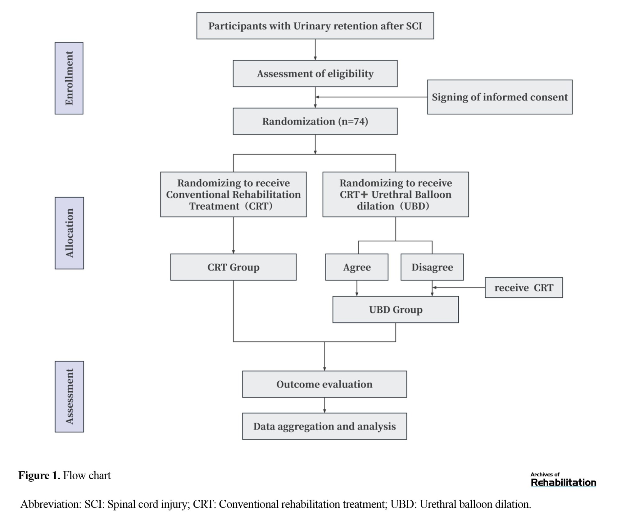
This study will be designed as a randomized controlled clinical trial adopting the Zelen methodology to evaluate the rehabilitative effects of urethral balloon dilation in patients with urinary retention following SCI. The trial will be carried out at Qilu Hospital of Shandong University and has obtained ethical approval from the Ethics Committee of Qilu Hospital, Shandong University (Approval No. KYLL-202503-011-1). In compliance with current ethical standards for clinical trials, key aspects of the study, including participant recruitment, allocation procedures, informed consent, intervention protocols, and data acquisition, are summarized in Figure 1.
Study participants
Participants are SCI patients with urinary retention hospitalized in the Department of Rehabilitation Medicine, Qilu Hospital of Shandong University. The entire trial process is illustrated in Figure 1, which describes the recruitment procedure for this study.
Eligibility criteria
Inclusion criteria
The inclusion criteria are as follows: 1) aged 16–80 years, 2) diagnosed with SCI (complete or incomplete) through CT or MRI imaging, meeting the diagnostic standards established in the 2019 International Standards for Neurological Classification of Spinal Cord Injury (ISNCSCI) by the American Spinal Injury Association (ASIA) [24]. 3) past the spinal shock phase, with clear consciousness and stable vital signs, 4) diagnosed with urinary retention, meeting clinical diagnostic standards (the patient had normal spontaneous urination before SCI, but after injury, exhibited urinary retention, including lower abdominal distension, bladder fullness, dull percussion sound, and bladder residual urine volume >300 mL as confirmed by bladder ultrasound, defined as the mean of three consecutive PVR measurements obtained within a 24-hour period. These patients also present with difficulty urinating [25], 5) urodynamic studies indicating a maximum urethral pressure >50 cm H₂O, 6) no evidence of tumors, benign prostatic hyperplasia (BPH), or other pathological conditions, 7) no indwelling catheterization, and 8) voluntary participation in the study with signed informed consent.
Exclusion criteria
Participants will be excluded if they meet any of the following conditions:
1) severe dysfunction of major organs (heart, brain, lungs) or mental and cognitive disorders, 2) electrolyte imbalance or acid-base disorders, 3) severe renal disease, history of bladder fistula surgery, or urethral sphincterotomy, and 4) presence of urinary tract infection at the time of medical evaluation.
Randomization and informed consent
This trial will adopt a Zelen design. First, after being fully informed about the purpose of the study, study procedures, and the potential benefits and risks of the two treatment strategies, all eligible patients will sign written informed consent for study participation and subsequent randomization. Thereafter, participants will be assigned (1:1) to the balloon dilation or standard-treatment group using block randomization, with allocation concealed by sealed opaque envelopes or an electronic randomization system. After randomization, patients allocated to the intervention arm (balloon dilation group) will receive additional, procedure-specific information about urethral balloon dilation and will be asked to sign a separate written consent for the procedure, including an explanation of potential benefits (e.g. improved voiding function) and risks (eg, urethral injury). Patients who agree will undergo urethral balloon dilation, whereas those who decline will instead receive conventional rehabilitation treatment. Patients allocated to the control arm(conventional rehabilitation group), having already provided trial-level informed consent, will receive standard conservative therapy according to the routine care pathway of our center. Their anonymized clinical data will be collected and analyzed for research purposes. In accordance with the intention-to-treat (ITT) principle for Zelen design trials, all participants will remain in their originally randomized group for the primary analysis, regardless of whether they accept or decline the intervention. Participants assigned to the intervention arm who decline procedure consent will receive conventional rehabilitation care but will remain included in the ITT analysis set.
The study protocol will be reviewed and approved by the Ethics Committee and will be conducted in accordance with the Declaration of Helsinki (World Medical Association, 2013 revision; with specific attention to the sections on ethics committee review and informed consent). Outcomes including urinary function, psychological status, and quality of life will be assessed in both groups to determine the efficacy and safety of urethral balloon dilation.
Blinding
Due to the distinctive procedural characteristics of ultrasound-guided urethral balloon dilatation, blinding patients and treating clinicians is not feasible. However, to minimize measurement bias, strict blinding procedures will be implemented for outcome assessors, data collectors, and statisticians. These personnel will remain unaware of group allocation and treatment details throughout the study to ensure objective and independent evaluation of all study outcomes.
Study interventions
Patients are instructed to adhere to a standardized hydration protocol with strict control over fluid intake.
Conventional rehabilitation treatment (control group)
Participants assigned to the control group will receive conventional rehabilitation care, including intermittent catheterization, acupuncture and pelvic floor muscle training. Intermittent catheterization will be guided by urodynamic examination results to develop a personalized drinking plan and determine the appropriate catheterization frequency based on the volume of PVR urine. Acupuncture treatment will target acupoints in the lower abdomen and lumbosacral region to improve urinary function, administered once daily, 5 days per week, for a total duration of 1 to 2 weeks. A pelvic floor therapist will assess the participant’s pelvic floor muscle condition and provide guidance on performing effective contraction and relaxation exercises to enhance pelvic floor muscle function. A fully standardized protocol for both acupuncture and therapist-assisted pelvic floor muscle training is provided in the Supplementary Appendix.
Urethral balloon dilation (intervention group)
Participants allocated to the intervention group will receive urethral balloon dilation in addition to conventional rehabilitation care. The procedures are as follows.
1) A 14-Fr urethral catheter will be filled with sterile saline to confirm balloon integrity; once integrity is verified, the saline will be fully withdrawn. 2) Ultrasound imaging will be used to precisely locate the external urethral sphincter. 3) The catheter will be fully lubricated and gently inserted into the urethra. 4) With real-time ultrasound guidance, 2–3.5 mL of sterile saline will be slowly instilled into the balloon to dilate the external urethral sphincter. 5) Patients will be instructed to attempt urination during the procedure, and any changes in voiding patterns will be monitored. 6) At the end of the procedure, the balloon will be completely deflated and the catheter will then be removed carefully.
This procedure will be performed 3 times per participant within 1-2 weeks. The initial inflation volume will be determined within this range according to urethral outlet resistance on baseline urodynamic testing. The inflation volume will increase by 0.5–1 mL at each session, guided by a predefined protocol that combines ultrasound assessment of balloon shape and sphincter opening with the patient’s subjective tolerance (tolerable distension without marked pain or discomfort and no urethral bleeding or signs of autonomic dysreflexia). Each session will last approximately 5 minutes and will be performed under ultrasound guidance by experienced physicians, with strict aseptic technique maintained throughout.
Study outcomes
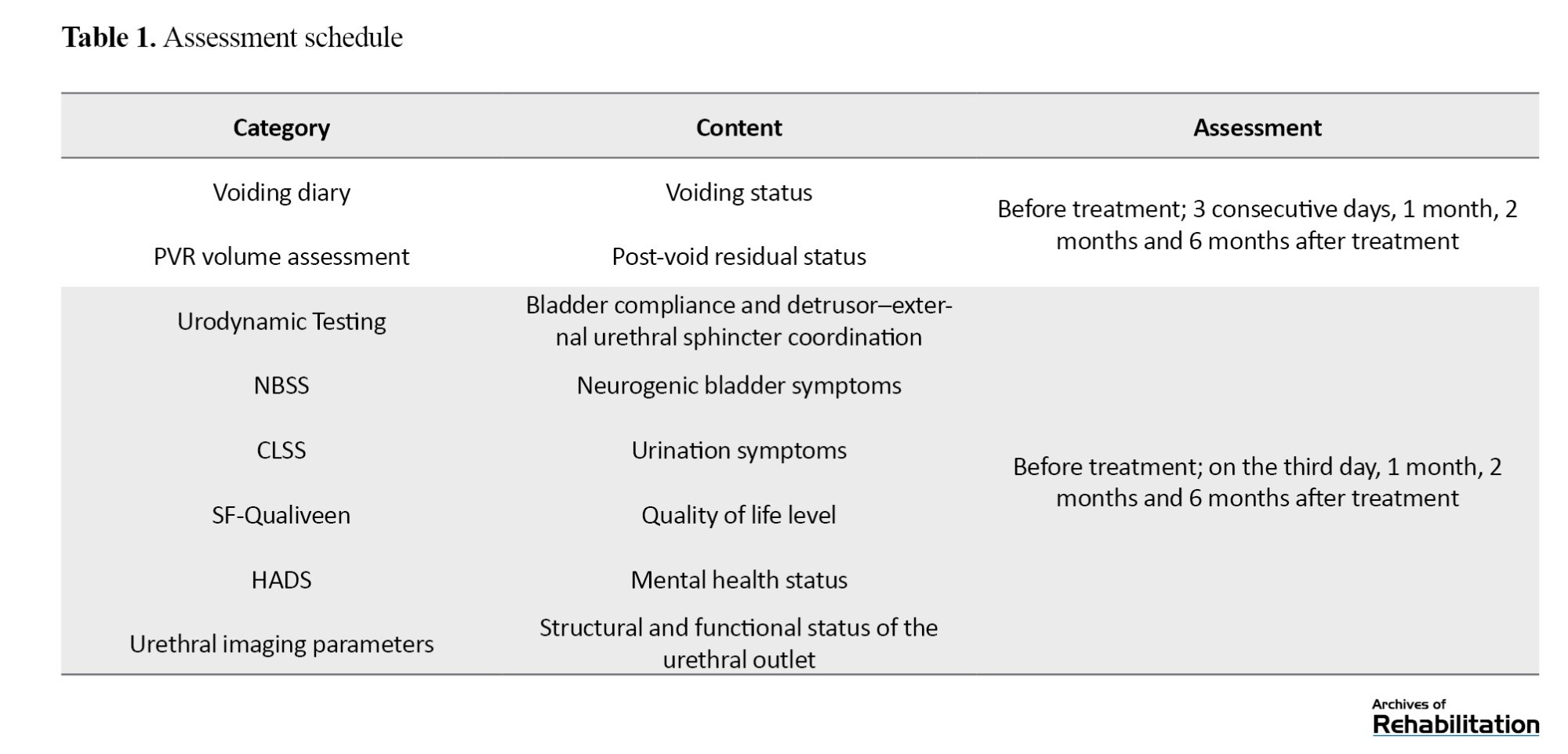
The assessment schedule is presented in Table 1.
Primary outcomes
PVR volume
PVR volume refers to the urine that remains in the bladder following voluntary voiding and is commonly used as a diagnostic tool. PVR measurement helps evaluate a range of conditions, such as neurogenic bladder, cauda equina syndrome, urinary outlet obstruction, mechanical obstruction, medication-related urinary retention, postoperative urinary retention, and urinary tract infection [26]. In this study, the HD5 bladder volume measurement device (produced by Hander Technology Co., Ltd., Liaoning, China) was used to assess PVR urine volume through real-time ultrasound imaging. For patients with spontaneous voiding ability, ultrasound was performed immediately after voiding to measure residual bladder volume. For patients who rely entirely on catheterization, bladder ultrasound was conducted prior to scheduled catheterization to record pre-catheterization bladder volume.
Voiding diary
A voiding diary will be provided to each participant, with instructions for consistent daily use. Before and after treatment, patients will record voiding patterns, including: daily spontaneous voided volume, catheterized urine volume, leakage volume and voiding frequency.
Urodynamic testing
Urodynamics is a standardized and quantitative assessment method that provides objective, physiologically based functional data. In this study, the Nidoc 970A urodynamic analyzer was used to perform standardized urodynamic testing. Key parameters monitored included maximum urinary flow rate (Qmax) and maximum urethral pressure (Pura max). Additionally, changes in urethral pressure distribution and detrusor–external urethral sphincter coordination were evaluated.
Secondary outcomes
Short-form health survey-qualiveen
The short-form health survey-qualiveen (SF-qualiveen) scale is designed to evaluate quality of life among patients with neurogenic bladder dysfunction. It evaluates 4 dimensions: Bother, limitations, fears, and feelings, with two questions per dimension. The scale was translated and validated in Chinese by Tang Rong, forming a neurogenic bladder-specific quality of life assessment tool. The Cronbach α coefficient for the full scale is 0.926, while the subscales range from 0.908 to 0.9, indicating high reliability and validity in assessing the quality of life in Chinese patients with neurogenic bladder [27, 28].
Neurogenic bladder symptom score (NBSS)
The NBSS comprises 24 items covering 3 key dimensions: urinary incontinence (8 items, 0–29 points), bladder storage and voiding function (7 items, 0–22 points), urinary tract complications (7 items, 0–23 points). Additionally, two non-scoring items assess bladder management methods and overall quality of life. Each item is rated on a 0–3 or 0–4 scale, with higher scores reflecting greater symptom severity. The Chinese version of NBSS has been validated for reliability and validity, making it a useful tool for assessing symptom improvement before and after intervention [29-31].
Core lower urinary tract symptom score (CLSS)
The CLSS will be applied to assess lower urinary tract symptoms before and after treatment. The total score ranges from 0 to 30, where 0 indicates no symptoms, 1 indicates rare symptoms, 2 indicates occasional symptoms, and 3 indicates frequent symptoms. Lower CLSS scores indicate greater symptom improvement following treatment [32, 33].
Hospital anxiety and depression scale (HADS)
HADS was developed by Zigmond and Snaith in 1983. It is used to screen for anxiety and depression in hospitalized patients [34]. It contains 14 items, comprising two subscales: 7 items for depression and 7 items for anxiety. The Chinese version of HADS has demonstrated strong reliability and validity, making it an effective tool for assessing psychological well-being in clinical settings [35].
Urethral imaging parameters
Urethral imaging parameters are utilized to quantitatively assess tissue changes before and after the intervention, providing data support for personalized treatment and enabling precise, visualized decision-making. The parameters measured included external urethral sphincter thickness, urethral diameter at the level of the sphincter, sphincter volume, and urethral mobility.
Before examination, participants are instructed to empty their bladder and bowels 30 minutes in advance, ensuring bladder volume <50 mL. Patients are placed in the lithotomy position in a supine posture. A LOGIQ e ultrasound machine with a 6–12 MHz high-frequency linear probe was used. The probe is placed longitudinally in the midline perineal area with the marker pointing upwards. Dynamic midsagittal images of the pelvic floor during Valsalva maneuvers are obtained. Each patient performed the Valsalva maneuver three times, and the best-quality image are selected for analysis.
Sphincter volume is calculated as the total volume of the urethral sphincter and enclosed urethra minus the urethral lumen volume. Volume is measured by outlining the contour of the target cross-section in successive 1 mm slices, and computed using the built-in ultrasound software formula (BK3dView software). Urethral segmental mobility is evaluated with urethral motion tracking software. For female patients, 6 equidistant points are marked from the bladder neck (Point 1) to the external urethral meatus (Point 6), dividing the urethra into 5 segments. For male patients, 6 equidistant landmarks are marked along the internal urethra from the bladder neck to the membranous urethra (including the prostatic and membranous parts). For each point, x and y coordinates relative to the inferior-posterior border of the pubic symphysis were recorded. Segmental mobility was calculated using the Equation 1: Quantifying the displacement vector for each segment [36, 37].

Follow-up
In addition to the immediate post-intervention evaluation, this study includes a structured follow-up schedule to assess the durability of treatment effects. All outcome measures, including PVR volume, voiding diary records, urodynamic parameters, NBSS, CLSS, quality of life (SF-qualiveen), and psychological status (HADS), will be reassessed at one month, two months and six months after completion of the three-session UBD intervention. These follow-up assessments will allow us to observe changes beyond the short-term treatment window and to determine whether improvements in bladder emptying, urinary symptoms, and quality of life are sustained over time.
Meanwhile, for hospitalized patients, the research team will conduct daily assessments to monitor the current clinical status, with a particular focus on post-intervention urethral injury and urinary function. All relevant data will be promptly recorded and analyzed to ensure real-time monitoring of the intervention’s effectiveness.
For discharged patients, a systematic follow-up mechanism has been established to maintain continuous communication between the patients and the medical team. A dedicated follow-up group chat will be created and managed by specialist rehabilitation physicians, rehabilitation therapists, and postgraduate rehabilitation researchers to provide timely, individualized assessments and guidance. Patients can consult the group chat at any time to ask questions related to their recovery. The medical team will offer professional advice and support based on the patient’s specific condition. Regular evaluations of patients, recovery progress will be conducted, and follow-up protocols will be adjusted accordingly based on patient feedback.
Study quality control and data management
This trial protocol has been reviewed and endorsed by experts in methodology and statistics. The study will be implemented in strict compliance with the approved protocol, and any protocol amendments will require approval from the Ethics Committee.
Training and standardization
Before the trial, all participating researchers will undergo standardized training to ensure familiarity with Zelen design methodology, urodynamic testing techniques, ultrasound-guided intervention techniques, urethral balloon dilation procedures, standardized follow-up protocols. Also, regular research meetings will be held to address potential issues encountered during the trial. Ultrasound guidance and balloon dilation procedures will be performed by experienced, specially trained professionals, following standardized protocols to minimize inter-operator variability. Outcome assessors, particularly those responsible for ultrasound measurements and urodynamic analyses, will receive dedicated calibration training, and inter-rater reliability for key quantitative measures will be evaluated in a subset of participants using intraclass correlation coefficients.
Data collection and security
All raw data will be systematically recorded in case report forms (CRFs), informed consent documents, and clinical records. All data will be fully traceable. Investigators will manually input CRF data, including their names and dates for accountability. Data collection will be conducted at regular intervals, and any modifications or missing data will be documented with detailed annotations.
Sample size calculation
The sample size for this trial was determined based on preliminary trial data from our center and general statistical considerations. In a pilot study conducted prior to this trial, patients with SCI-related urinary retention who met the present eligibility criteria were allocated to receive either UBD or conventional rehabilitation. In this preliminary dataset, the mean PVR after treatment was 385 mL in the conventional rehabilitation group and 331 mL in the UBD group, with a pooled standard deviation of approximately 67 mL. Assuming a two-sided α of 0.05, a power of 1–β=0.90 (90%), and an equal allocation ratio (1:1), a sample size calculation tool indicated that 33 patients per group would be required. Allowing for an anticipated dropout rate of about 10%, the final planned sample size was 74 patients, with 37 in the intervention group and 37 in the control group.
Statistical analysis
Data will be independently entered into Excel by two investigators, with subsequent cross-checking to ensure consistency. Statistical analyses will be conducted using SPSS software, version 26.0. Unless otherwise stated, two-sided tests will be applied, and a P<0.05 will be considered statistically significant.
Analysis populations
The primary efficacy analysis will be conducted under the ITT principle and will include all randomized participants, analyzed according to their originally assigned group (UBD group and conventional rehabilitation group), regardless of actual treatment received, treatment adherence, or subsequent withdrawal.
The Per-protocol PP set will include participants who complete the allocated treatment and the primary endpoint assessment without major protocol deviations (e.g. substantial non-compliance, receiving additional non-protocol interventions that could affect voiding function). Key efficacy analyses will be repeated in the PP set as sensitivity analyses.
Descriptive statistics
Baseline demographic and clinical characteristics will be summarized separately for each group. Continuous variables will be described using Mean±SD if approximately normally distributed, or median and interquartile range (IQR) otherwise. Categorical variables will be presented as counts and percentages.
Primary outcome analysis
The primary outcome of this study is PVR measured at prespecified time points, with PVR at the end of the treatment period serving as the primary assessment time point. PVR will also be measured at 1, 2 and 6 months after the intervention to characterize its longitudinal trajectory . For continuous variables, distribution normality will be evaluated using the Shapiro-Wilk test. For the primary comparison, the mean difference in PVR at the end of treatment between the two groups will be examined. If PVR values at this time point are approximately normally distributed, an independent samples t-test will be applied; otherwise, the Mann-Whitney U test will be used. In addition, a change in PVR from baseline to the end of treatment will be calculated, and the between-group differences in mean change will be analyzed using the same statistical approach. To fully use the repeated measurements over time (end of treatment, 1 month, 2 months, and 6 months after), a repeated-measures approach, including repeated-measures analysis of variance (ANOVA) or a linear mixed-effects model (with group, time, and group-by-time interaction as fixed effects and participant-level random intercepts), will be performed as a supplementary analysis. Baseline PVR and important prognostic factors (e.g. neurological injury level and injury completeness) may be included as covariates in adjusted models if they improve model fit.
Secondary outcome analysis
Continuous secondary outcomes (e.g. other urodynamic parameters, voiding diary variables, NBSS, CLSS, SF-qualiveen and HADS scores, ultrasound parameters) will be analyzed using methods similar to those for the primary outcome. For single post-treatment time points, between-group comparisons will be performed using independent samples t-tests or Mann-Whitney U tests depending on the data distribution. For outcomes assessed repeatedly over time, repeated-measures ANOVA or linear mixed-effects models will be used, with group, time and group-by-time interaction specified as fixed effects. Where necessary, data transformations or appropriate generalized linear models will be used for clearly non-normal distributions. Categorical outcomes, including adverse events incidence and the proportion of patients achieving predefined clinically meaningful improvement (e.g. a specified reduction in PVR), will be compared between groups using the chi-squared tests or Fisher exact tests as appropriate. When covariate adjustment is required, logistic regression models will be applied, and effect estimates will be reported as odds ratios with 95% confidence intervals.
Handling of missing data
The extent and pattern of missing data will be summarized for each outcome by treatment group and assessment time point. For the primary endpoint, if the proportion of missing data is ≤5%, the main ITT analysis will be based on complete cases, accompanied by a sensitivity analysis using multiple imputation. If missing data exceed 5%, multiple imputation using chained equations will be used for the primary ITT analysis. The imputation model will include treatment group, baseline values of the outcome, neurological injury level (cervical/thoracic/lumbar), injury completeness (complete/incomplete) and other key prognostic variables. Results from analyses using imputed datasets and complete cases will be compared to evaluate the robustness of the conclusions. The same approach to missing data will be applied to secondary outcomes when appropriate.
Additional analyses (subgroup and sensitivity analyses)
To explore the potential impact of patient heterogeneity, exploratory subgroup analyses of the primary outcome will be performed according to neurological injury level (cervical, thoracic, or lumbar) and injury completeness (complete or incomplete) by including corresponding interaction terms (group × subgroup) in the relevant models. If there is clinical interest, sex and time since injury (e.g. ≤6 months or >6 months) may also be explored. Sensitivity analyses will include: repeating the primary analysis in the PP population; and repeating the ITT analysis after excluding participants randomized to the UBD group who ultimately refuse the UBD procedure.
Discussion
Based on the guidelines for the urological management and clinical rehabilitation of SCI patients, the long-term management goals for SCI-related neurogenic lower urinary tract dysfunction are categorized into primary and secondary objectives [38]. The primary objective is to preserve upper urinary tract function, particularly renal function, by keeping bladder pressure within a safe range during both the storage and voiding phases. Secondary objective include partially or fully restoring lower urinary tract function, improving urinary continence, reducing PVR urine, preventing urinary tract infections, and thereby enhancing the patient’s quality of life [39].
At present, commonly used approaches for urinary retention after SCI include indwelling catheterization, clean intermittent catheterization, bladder training, pharmacotherapy, and electrical stimulation therapy. Although these approaches can alleviate urinary retention to some extent, each has certain limitations. Indwelling catheterization allows continuous drainage and relieves acute symptoms, but prolonged use carries higher risks of urinary tract infections, urethral injury, and catheter dependence, which may hinder the reestablishment of normal voiding reflexes. Intermittent catheterization is a key strategy for managing neurogenic bladder in patients with SCI, as it may lower the likelihood of urinary tract infections and help preserve bladder function. However, it requires strict aseptic technique and a high level of adherence from patients or caregivers [10, 40, 41]. Inadequate mastery of catheterization techniques, inappropriate catheter selection, or insufficient lubrication may lead to urethral irritation or mucosal injury, thereby increasing the risk of infection. In addition, the need for multiple catheterizations each day and the associated long-term cost of supplies can create inconvenience and financial burden, which may negatively affect treatment adherence [42]. Bladder training is applicable to some patients with partial neural preservation but is often slow in progress and varies greatly among individuals. Pharmacotherapy may improve bladder function, but it is often accompanied by adverse effects, including dry mouth, constipation, and hypotension [43-45]. Electrical stimulation treatments (eg, sacral neuromodulation, transcutaneous electrical stimulation) may support functional recovery but are costly, time-consuming, and not suitable for all patients [46]. Therefore, urinary retention following SCI remains an important clinical challenge.
This study proposes a structured and standardized protocol for ultrasound-guided UBD, offering a procedural approach that may help reduce urethral outlet resistance, facilitate more effective bladder emptying, and decrease long-term catheter dependence. This protocol therefore proposes a clinically feasible and research-worthy intervention option for the rehabilitation management of urinary retention after SCI. As a non-surgical physical approach, UBD has potential clinical value and practical advantages for addressing urinary retention in this population. This technique is minimally invasive and easy to perform, involving the mechanical expansion of urethral strictures via the insertion of a balloon catheter, without the need for surgical incisions or anesthesia, making it well-tolerated by patients. The procedure is gentle and helps minimize postoperative complications and discomfort caused by the intervention. Under ultrasound guidance, UBD enables precise localization and real-time monitoring of the urethral stricture, thereby ensuring the accuracy of the dilation. These factors contribute to greater patient acceptance and adherence [43, 44], enhancing the potential for broader clinical implementation of this intervention.
In this study, ultrasound imaging parameters will be used to quantitatively evaluate structural changes in the urethra before and after intervention, including measurements of external urethral sphincter thickness, urethral diameter at the sphincter region, and sphincter volume, to accurately reflect the anatomical impact and effectiveness of balloon dilatation. Additionally, for the first time, “urethral mobility” will be introduced as an exploratory imaging parameter into the assessment framework of neurogenic bladder, serving as an imaging-based representation of the mobility of the urethra and adjacent tissues [47]. In prior studies, urethral mobility has been used mainly to evaluate urethral and bladder neck support and dynamic stability in women with stress urinary incontinence, where it has shown potential clinical relevance in this population [37, 48]. We hypothesize that under the pathological conditions of SCI—particularly in patients with elevated external sphincter tone or increased urethral outlet resistance—urethral mobility may closely correlate with the flexibility and relaxation capacity of sphincteric muscle tissues. Assessing urethral mobility could reflect the tissue’s deformation capacity under stress and serve as an indirect indicator of local mechanical compliance. By examining segmental urethral displacement patterns before and after intervention, this study seeks to explore the potential utility of urethral mobility as a functional imaging marker for evaluating changes in urethral dynamics, and to provide preliminary data for future mechanistic studies and clinical assessment systems.
This study adopted a randomized controlled design based on Zelen approach. After informed consent was obtained and randomization was completed, participants allocated to the experimental group were allowed to make an autonomous decision regarding UBD as a new treatment option once they had received sufficient information, which more closely reflects real decision-making processes in rehabilitation practice. The purpose of this trial is not merely to assess the ideal efficacy of UBD among patients randomized to the UBD group who fully adhere to the intervention, but rather to capture the overall real-world effect of offering UBD as a treatment option on top of standard rehabilitation for eligible SCI patients. Under the premise that all participants provide informed consent and that their treatment preferences are fully respected, we believe that this design may enhance the study’ s external validity and increase its applicability to clinical decision-making [49, 50]. It should be noted that some participants randomized to the UBD group may ultimately not receive UBD, which may lead to a somewhat conservative estimate of the treatment effect in ITT analyses. To more comprehensively characterize the intervention effect, the protocol prespecifies per-protocol analyses and sensitivity analyses as important complementary approaches.
This study will have several limitations. It is a single-center trial conducted in a Grade A tertiary hospital in China, and the characteristics of the enrolled patients and local rehabilitation practice in this setting may not fully represent those in other institutions, regions or healthcare systems. Because patient blinding is not feasible for this procedure-based intervention, expectancy effects may influence patient-reported outcomes; however, our primary endpoints are largely objective measures (ultrasound-measured PVR and standardized urodynamic parameters) and are therefore less susceptible to such bias. In addition, the relatively modest sample size may, to some extent, affect the precision of effect estimates and the generalizability of the results. Therefore, future multicenter studies with larger sample sizes in more diverse settings are warranted to further confirm the robustness of the findings and enhance their external validity.
Conclusion
This study protocol describes a randomized controlled trial designed to evaluate whether ultrasound-guided UBD may help improve urinary retention and reduce catheter dependence in patients with SCI. The evidence generated from this study is expected to inform and optimize clinical rehabilitation strategies for neurogenic bladder management. In the future, based on the findings of this study, we plan to conduct larger-scale, multicenter clinical trials with extended follow-up periods to further validate the efficacy, safety, and long-term durability of UBD. Such efforts will enhance the clinical applicability and generalizability of the results.
Ethical Considerations
Compliance with ethical guidelines
This study was approved by the Ethics Committee of Qilu Hospital, Shandong University, Jinan, China (Code: KYLL-202503-011-1).
Funding
This research did not receive any specific grant from funding agencies in the public, commercial, or not-for-profit sectors.
Authors' contributions
Investigation: Fuchao Yao, Xiaomin Lu and Yirong Zha; Formal analysis and data collection: Fuchao Yao, Xiaomin Lu and Jinlu Li; Conceptualization: Fuchao Yao and Hui Wei; Validation: Xiaomin Lu; Writing the original draft: Fuchao Yao; Review, and editing: Xiaomin Lu, Hui Wei and Jinlu Li; Methodology: Fuchao Yao, Hui Wei and Jinlu Li; Visualization: Jinlu Li; Supervision: Hui Wei.
Conflict of interest
The authors declared no conflict of interest.
Acknowledgments
The authors gratefully acknowledge the support provided by the Department of Rehabilitation Medicine, Qilu Hospital of Shandong University.
References
Spinal cord injury (SCI) frequently leads to lower urinary tract dysfunction, with urinary retention being one of the most common and detrimental manifestations. After SCI, normal voiding becomes impaired, and patients may also present with elevated urethral outlet resistance [1, 2]. This condition leads to ineffective voiding, increased post-void residual (PVR) volume, and elevated outlet resistance, which in turn can cause increased intravesical pressure, urinary tract infections, vesicoureteral reflux, upper urinary tract damage, and impaired renal function [3-5]—thereby adversely affecting patients’ overall well-being and longer-term clinical outcomes. In addition to physiological complications, urinary retention and related voiding disorders often impose a substantial psychological burden. Studies have shown that patients who are dependent on catheterization or suffer recurrent urinary complications are more prone to anxiety, depression, social withdrawal, and diminished self-esteem, further reducing treatment adherence and life satisfaction. The interplay of physical and psychological stressors substantially compromises the overall health status of individuals with SCI [6-8]. Previous studies have identified urinary retention as a major contributor to late-stage mortality in this population [2, 9-11]. Therefore, accurate assessment and effective intervention for urinary retention have become urgent priorities in both clinical and rehabilitation settings.
Balloon dilatation is a well-established physical intervention that has demonstrated favorable safety and efficacy profiles in various clinical conditions [12-18]. By mechanically expanding narrowed or obstructed tissues, it helps to restore the anatomical structure and functional patency of affected pathways. When applied to muscular tissue, balloon dilatation can effectively enhance local tissue compliance and coordination, improving both relaxation and contractile capabilities [14, 19]. The technique is characterized by its simplicity, low complication rate, good patient tolerability, and cost-effectiveness, making it a highly feasible option for broader clinical application [10, 20]. Given the critical role of elevated external urethral sphincter tone in the pathophysiology of urinary retention following SCI, this study applies balloon dilatation to reduce sphincteric tension and this approach may offer a novel and promising treatment pathway for improving voiding function in SCI-related urinary retention. Therefore, a systematic exploration of its application in this patient population holds significant clinical relevance and practical value.
Ultrasound, as a medical imaging technique based on high-frequency sound waves, offers notable advantages, such as non-invasiveness, absence of radiation, operational simplicity, real-time imaging, and repeatability [21, 22]. It is commonly applied to assess both anatomical characteristics and functional performance of the urinary tract. In clinical settings, ultrasound provides clear imaging of the urethra and bladder, as well as adjacent soft tissues, and allows dynamic evaluation of tissue motion during physiological processes. These features make it particularly suitable for research and interventional guidance related to voiding function, and highly acceptable and safe in the diagnosis and management of neurogenic bladder [23]. In this study, ultrasound will be employed to guide the urethral balloon dilatation (UBD) procedure. Real-time imaging facilitates accurate localization of the external urethral sphincter and assessment of the dilatation range, thereby enhancing the precision of the intervention and reducing the risk of tissue injury. Furthermore, ultrasound also supports dynamic evaluation of treatment efficacy. Through visualizing structural and functional changes, such as external urethral sphincter thickness, urethral diameter at the sphincter level, sphincter volume, and urethral mobility, ultrasound provides valuable imaging-based support for both therapeutic assessment and mechanistic exploration.
Therefore, this study focuses on the therapeutic potential of ultrasound-guided UBD in patients with urinary retention following SCI. By leveraging real-time imaging, the procedure enables precise control of balloon positioning and dilatation range, thereby enhancing the safety and controllability of the intervention. The study aims to systematically evaluate the clinical effectiveness of this technique in improving voiding function, alleviating functional obstruction, and protecting upper urinary tract integrity, while exploring its broader application value in the management of neurogenic bladder. This minimally invasive approach is expected to offer a new clinical pathway for individualized treatment of urinary retention following SCI.
Materials and Methods
Study design
This study will be designed as a randomized controlled clinical trial adopting the Zelen methodology to evaluate the rehabilitative effects of urethral balloon dilation in patients with urinary retention following SCI. The trial will be carried out at Qilu Hospital of Shandong University and has obtained ethical approval from the Ethics Committee of Qilu Hospital, Shandong University (Approval No. KYLL-202503-011-1). In compliance with current ethical standards for clinical trials, key aspects of the study, including participant recruitment, allocation procedures, informed consent, intervention protocols, and data acquisition, are summarized in Figure 1.
Study participants
Participants are SCI patients with urinary retention hospitalized in the Department of Rehabilitation Medicine, Qilu Hospital of Shandong University. The entire trial process is illustrated in Figure 1, which describes the recruitment procedure for this study.
Eligibility criteria
Inclusion criteria
The inclusion criteria are as follows: 1) aged 16–80 years, 2) diagnosed with SCI (complete or incomplete) through CT or MRI imaging, meeting the diagnostic standards established in the 2019 International Standards for Neurological Classification of Spinal Cord Injury (ISNCSCI) by the American Spinal Injury Association (ASIA) [24]. 3) past the spinal shock phase, with clear consciousness and stable vital signs, 4) diagnosed with urinary retention, meeting clinical diagnostic standards (the patient had normal spontaneous urination before SCI, but after injury, exhibited urinary retention, including lower abdominal distension, bladder fullness, dull percussion sound, and bladder residual urine volume >300 mL as confirmed by bladder ultrasound, defined as the mean of three consecutive PVR measurements obtained within a 24-hour period. These patients also present with difficulty urinating [25], 5) urodynamic studies indicating a maximum urethral pressure >50 cm H₂O, 6) no evidence of tumors, benign prostatic hyperplasia (BPH), or other pathological conditions, 7) no indwelling catheterization, and 8) voluntary participation in the study with signed informed consent.
Exclusion criteria
Participants will be excluded if they meet any of the following conditions:
1) severe dysfunction of major organs (heart, brain, lungs) or mental and cognitive disorders, 2) electrolyte imbalance or acid-base disorders, 3) severe renal disease, history of bladder fistula surgery, or urethral sphincterotomy, and 4) presence of urinary tract infection at the time of medical evaluation.
Randomization and informed consent
This trial will adopt a Zelen design. First, after being fully informed about the purpose of the study, study procedures, and the potential benefits and risks of the two treatment strategies, all eligible patients will sign written informed consent for study participation and subsequent randomization. Thereafter, participants will be assigned (1:1) to the balloon dilation or standard-treatment group using block randomization, with allocation concealed by sealed opaque envelopes or an electronic randomization system. After randomization, patients allocated to the intervention arm (balloon dilation group) will receive additional, procedure-specific information about urethral balloon dilation and will be asked to sign a separate written consent for the procedure, including an explanation of potential benefits (e.g. improved voiding function) and risks (eg, urethral injury). Patients who agree will undergo urethral balloon dilation, whereas those who decline will instead receive conventional rehabilitation treatment. Patients allocated to the control arm(conventional rehabilitation group), having already provided trial-level informed consent, will receive standard conservative therapy according to the routine care pathway of our center. Their anonymized clinical data will be collected and analyzed for research purposes. In accordance with the intention-to-treat (ITT) principle for Zelen design trials, all participants will remain in their originally randomized group for the primary analysis, regardless of whether they accept or decline the intervention. Participants assigned to the intervention arm who decline procedure consent will receive conventional rehabilitation care but will remain included in the ITT analysis set.
The study protocol will be reviewed and approved by the Ethics Committee and will be conducted in accordance with the Declaration of Helsinki (World Medical Association, 2013 revision; with specific attention to the sections on ethics committee review and informed consent). Outcomes including urinary function, psychological status, and quality of life will be assessed in both groups to determine the efficacy and safety of urethral balloon dilation.
Blinding
Due to the distinctive procedural characteristics of ultrasound-guided urethral balloon dilatation, blinding patients and treating clinicians is not feasible. However, to minimize measurement bias, strict blinding procedures will be implemented for outcome assessors, data collectors, and statisticians. These personnel will remain unaware of group allocation and treatment details throughout the study to ensure objective and independent evaluation of all study outcomes.
Study interventions
Patients are instructed to adhere to a standardized hydration protocol with strict control over fluid intake.
Conventional rehabilitation treatment (control group)
Participants assigned to the control group will receive conventional rehabilitation care, including intermittent catheterization, acupuncture and pelvic floor muscle training. Intermittent catheterization will be guided by urodynamic examination results to develop a personalized drinking plan and determine the appropriate catheterization frequency based on the volume of PVR urine. Acupuncture treatment will target acupoints in the lower abdomen and lumbosacral region to improve urinary function, administered once daily, 5 days per week, for a total duration of 1 to 2 weeks. A pelvic floor therapist will assess the participant’s pelvic floor muscle condition and provide guidance on performing effective contraction and relaxation exercises to enhance pelvic floor muscle function. A fully standardized protocol for both acupuncture and therapist-assisted pelvic floor muscle training is provided in the Supplementary Appendix.
Urethral balloon dilation (intervention group)
Participants allocated to the intervention group will receive urethral balloon dilation in addition to conventional rehabilitation care. The procedures are as follows.
1) A 14-Fr urethral catheter will be filled with sterile saline to confirm balloon integrity; once integrity is verified, the saline will be fully withdrawn. 2) Ultrasound imaging will be used to precisely locate the external urethral sphincter. 3) The catheter will be fully lubricated and gently inserted into the urethra. 4) With real-time ultrasound guidance, 2–3.5 mL of sterile saline will be slowly instilled into the balloon to dilate the external urethral sphincter. 5) Patients will be instructed to attempt urination during the procedure, and any changes in voiding patterns will be monitored. 6) At the end of the procedure, the balloon will be completely deflated and the catheter will then be removed carefully.
This procedure will be performed 3 times per participant within 1-2 weeks. The initial inflation volume will be determined within this range according to urethral outlet resistance on baseline urodynamic testing. The inflation volume will increase by 0.5–1 mL at each session, guided by a predefined protocol that combines ultrasound assessment of balloon shape and sphincter opening with the patient’s subjective tolerance (tolerable distension without marked pain or discomfort and no urethral bleeding or signs of autonomic dysreflexia). Each session will last approximately 5 minutes and will be performed under ultrasound guidance by experienced physicians, with strict aseptic technique maintained throughout.
Study outcomes
The assessment schedule is presented in Table 1.
Primary outcomes
PVR volume
PVR volume refers to the urine that remains in the bladder following voluntary voiding and is commonly used as a diagnostic tool. PVR measurement helps evaluate a range of conditions, such as neurogenic bladder, cauda equina syndrome, urinary outlet obstruction, mechanical obstruction, medication-related urinary retention, postoperative urinary retention, and urinary tract infection [26]. In this study, the HD5 bladder volume measurement device (produced by Hander Technology Co., Ltd., Liaoning, China) was used to assess PVR urine volume through real-time ultrasound imaging. For patients with spontaneous voiding ability, ultrasound was performed immediately after voiding to measure residual bladder volume. For patients who rely entirely on catheterization, bladder ultrasound was conducted prior to scheduled catheterization to record pre-catheterization bladder volume.
Voiding diary
A voiding diary will be provided to each participant, with instructions for consistent daily use. Before and after treatment, patients will record voiding patterns, including: daily spontaneous voided volume, catheterized urine volume, leakage volume and voiding frequency.
Urodynamic testing
Urodynamics is a standardized and quantitative assessment method that provides objective, physiologically based functional data. In this study, the Nidoc 970A urodynamic analyzer was used to perform standardized urodynamic testing. Key parameters monitored included maximum urinary flow rate (Qmax) and maximum urethral pressure (Pura max). Additionally, changes in urethral pressure distribution and detrusor–external urethral sphincter coordination were evaluated.
Secondary outcomes
Short-form health survey-qualiveen
The short-form health survey-qualiveen (SF-qualiveen) scale is designed to evaluate quality of life among patients with neurogenic bladder dysfunction. It evaluates 4 dimensions: Bother, limitations, fears, and feelings, with two questions per dimension. The scale was translated and validated in Chinese by Tang Rong, forming a neurogenic bladder-specific quality of life assessment tool. The Cronbach α coefficient for the full scale is 0.926, while the subscales range from 0.908 to 0.9, indicating high reliability and validity in assessing the quality of life in Chinese patients with neurogenic bladder [27, 28].
Neurogenic bladder symptom score (NBSS)
The NBSS comprises 24 items covering 3 key dimensions: urinary incontinence (8 items, 0–29 points), bladder storage and voiding function (7 items, 0–22 points), urinary tract complications (7 items, 0–23 points). Additionally, two non-scoring items assess bladder management methods and overall quality of life. Each item is rated on a 0–3 or 0–4 scale, with higher scores reflecting greater symptom severity. The Chinese version of NBSS has been validated for reliability and validity, making it a useful tool for assessing symptom improvement before and after intervention [29-31].
Core lower urinary tract symptom score (CLSS)
The CLSS will be applied to assess lower urinary tract symptoms before and after treatment. The total score ranges from 0 to 30, where 0 indicates no symptoms, 1 indicates rare symptoms, 2 indicates occasional symptoms, and 3 indicates frequent symptoms. Lower CLSS scores indicate greater symptom improvement following treatment [32, 33].
Hospital anxiety and depression scale (HADS)
HADS was developed by Zigmond and Snaith in 1983. It is used to screen for anxiety and depression in hospitalized patients [34]. It contains 14 items, comprising two subscales: 7 items for depression and 7 items for anxiety. The Chinese version of HADS has demonstrated strong reliability and validity, making it an effective tool for assessing psychological well-being in clinical settings [35].
Urethral imaging parameters
Urethral imaging parameters are utilized to quantitatively assess tissue changes before and after the intervention, providing data support for personalized treatment and enabling precise, visualized decision-making. The parameters measured included external urethral sphincter thickness, urethral diameter at the level of the sphincter, sphincter volume, and urethral mobility.
Before examination, participants are instructed to empty their bladder and bowels 30 minutes in advance, ensuring bladder volume <50 mL. Patients are placed in the lithotomy position in a supine posture. A LOGIQ e ultrasound machine with a 6–12 MHz high-frequency linear probe was used. The probe is placed longitudinally in the midline perineal area with the marker pointing upwards. Dynamic midsagittal images of the pelvic floor during Valsalva maneuvers are obtained. Each patient performed the Valsalva maneuver three times, and the best-quality image are selected for analysis.
Sphincter volume is calculated as the total volume of the urethral sphincter and enclosed urethra minus the urethral lumen volume. Volume is measured by outlining the contour of the target cross-section in successive 1 mm slices, and computed using the built-in ultrasound software formula (BK3dView software). Urethral segmental mobility is evaluated with urethral motion tracking software. For female patients, 6 equidistant points are marked from the bladder neck (Point 1) to the external urethral meatus (Point 6), dividing the urethra into 5 segments. For male patients, 6 equidistant landmarks are marked along the internal urethra from the bladder neck to the membranous urethra (including the prostatic and membranous parts). For each point, x and y coordinates relative to the inferior-posterior border of the pubic symphysis were recorded. Segmental mobility was calculated using the Equation 1: Quantifying the displacement vector for each segment [36, 37].
Follow-up
In addition to the immediate post-intervention evaluation, this study includes a structured follow-up schedule to assess the durability of treatment effects. All outcome measures, including PVR volume, voiding diary records, urodynamic parameters, NBSS, CLSS, quality of life (SF-qualiveen), and psychological status (HADS), will be reassessed at one month, two months and six months after completion of the three-session UBD intervention. These follow-up assessments will allow us to observe changes beyond the short-term treatment window and to determine whether improvements in bladder emptying, urinary symptoms, and quality of life are sustained over time.
Meanwhile, for hospitalized patients, the research team will conduct daily assessments to monitor the current clinical status, with a particular focus on post-intervention urethral injury and urinary function. All relevant data will be promptly recorded and analyzed to ensure real-time monitoring of the intervention’s effectiveness.
For discharged patients, a systematic follow-up mechanism has been established to maintain continuous communication between the patients and the medical team. A dedicated follow-up group chat will be created and managed by specialist rehabilitation physicians, rehabilitation therapists, and postgraduate rehabilitation researchers to provide timely, individualized assessments and guidance. Patients can consult the group chat at any time to ask questions related to their recovery. The medical team will offer professional advice and support based on the patient’s specific condition. Regular evaluations of patients, recovery progress will be conducted, and follow-up protocols will be adjusted accordingly based on patient feedback.
Study quality control and data management
This trial protocol has been reviewed and endorsed by experts in methodology and statistics. The study will be implemented in strict compliance with the approved protocol, and any protocol amendments will require approval from the Ethics Committee.
Training and standardization
Before the trial, all participating researchers will undergo standardized training to ensure familiarity with Zelen design methodology, urodynamic testing techniques, ultrasound-guided intervention techniques, urethral balloon dilation procedures, standardized follow-up protocols. Also, regular research meetings will be held to address potential issues encountered during the trial. Ultrasound guidance and balloon dilation procedures will be performed by experienced, specially trained professionals, following standardized protocols to minimize inter-operator variability. Outcome assessors, particularly those responsible for ultrasound measurements and urodynamic analyses, will receive dedicated calibration training, and inter-rater reliability for key quantitative measures will be evaluated in a subset of participants using intraclass correlation coefficients.
Data collection and security
All raw data will be systematically recorded in case report forms (CRFs), informed consent documents, and clinical records. All data will be fully traceable. Investigators will manually input CRF data, including their names and dates for accountability. Data collection will be conducted at regular intervals, and any modifications or missing data will be documented with detailed annotations.
Sample size calculation
The sample size for this trial was determined based on preliminary trial data from our center and general statistical considerations. In a pilot study conducted prior to this trial, patients with SCI-related urinary retention who met the present eligibility criteria were allocated to receive either UBD or conventional rehabilitation. In this preliminary dataset, the mean PVR after treatment was 385 mL in the conventional rehabilitation group and 331 mL in the UBD group, with a pooled standard deviation of approximately 67 mL. Assuming a two-sided α of 0.05, a power of 1–β=0.90 (90%), and an equal allocation ratio (1:1), a sample size calculation tool indicated that 33 patients per group would be required. Allowing for an anticipated dropout rate of about 10%, the final planned sample size was 74 patients, with 37 in the intervention group and 37 in the control group.
Statistical analysis
Data will be independently entered into Excel by two investigators, with subsequent cross-checking to ensure consistency. Statistical analyses will be conducted using SPSS software, version 26.0. Unless otherwise stated, two-sided tests will be applied, and a P<0.05 will be considered statistically significant.
Analysis populations
The primary efficacy analysis will be conducted under the ITT principle and will include all randomized participants, analyzed according to their originally assigned group (UBD group and conventional rehabilitation group), regardless of actual treatment received, treatment adherence, or subsequent withdrawal.
The Per-protocol PP set will include participants who complete the allocated treatment and the primary endpoint assessment without major protocol deviations (e.g. substantial non-compliance, receiving additional non-protocol interventions that could affect voiding function). Key efficacy analyses will be repeated in the PP set as sensitivity analyses.
Descriptive statistics
Baseline demographic and clinical characteristics will be summarized separately for each group. Continuous variables will be described using Mean±SD if approximately normally distributed, or median and interquartile range (IQR) otherwise. Categorical variables will be presented as counts and percentages.
Primary outcome analysis
The primary outcome of this study is PVR measured at prespecified time points, with PVR at the end of the treatment period serving as the primary assessment time point. PVR will also be measured at 1, 2 and 6 months after the intervention to characterize its longitudinal trajectory . For continuous variables, distribution normality will be evaluated using the Shapiro-Wilk test. For the primary comparison, the mean difference in PVR at the end of treatment between the two groups will be examined. If PVR values at this time point are approximately normally distributed, an independent samples t-test will be applied; otherwise, the Mann-Whitney U test will be used. In addition, a change in PVR from baseline to the end of treatment will be calculated, and the between-group differences in mean change will be analyzed using the same statistical approach. To fully use the repeated measurements over time (end of treatment, 1 month, 2 months, and 6 months after), a repeated-measures approach, including repeated-measures analysis of variance (ANOVA) or a linear mixed-effects model (with group, time, and group-by-time interaction as fixed effects and participant-level random intercepts), will be performed as a supplementary analysis. Baseline PVR and important prognostic factors (e.g. neurological injury level and injury completeness) may be included as covariates in adjusted models if they improve model fit.
Secondary outcome analysis
Continuous secondary outcomes (e.g. other urodynamic parameters, voiding diary variables, NBSS, CLSS, SF-qualiveen and HADS scores, ultrasound parameters) will be analyzed using methods similar to those for the primary outcome. For single post-treatment time points, between-group comparisons will be performed using independent samples t-tests or Mann-Whitney U tests depending on the data distribution. For outcomes assessed repeatedly over time, repeated-measures ANOVA or linear mixed-effects models will be used, with group, time and group-by-time interaction specified as fixed effects. Where necessary, data transformations or appropriate generalized linear models will be used for clearly non-normal distributions. Categorical outcomes, including adverse events incidence and the proportion of patients achieving predefined clinically meaningful improvement (e.g. a specified reduction in PVR), will be compared between groups using the chi-squared tests or Fisher exact tests as appropriate. When covariate adjustment is required, logistic regression models will be applied, and effect estimates will be reported as odds ratios with 95% confidence intervals.
Handling of missing data
The extent and pattern of missing data will be summarized for each outcome by treatment group and assessment time point. For the primary endpoint, if the proportion of missing data is ≤5%, the main ITT analysis will be based on complete cases, accompanied by a sensitivity analysis using multiple imputation. If missing data exceed 5%, multiple imputation using chained equations will be used for the primary ITT analysis. The imputation model will include treatment group, baseline values of the outcome, neurological injury level (cervical/thoracic/lumbar), injury completeness (complete/incomplete) and other key prognostic variables. Results from analyses using imputed datasets and complete cases will be compared to evaluate the robustness of the conclusions. The same approach to missing data will be applied to secondary outcomes when appropriate.
Additional analyses (subgroup and sensitivity analyses)
To explore the potential impact of patient heterogeneity, exploratory subgroup analyses of the primary outcome will be performed according to neurological injury level (cervical, thoracic, or lumbar) and injury completeness (complete or incomplete) by including corresponding interaction terms (group × subgroup) in the relevant models. If there is clinical interest, sex and time since injury (e.g. ≤6 months or >6 months) may also be explored. Sensitivity analyses will include: repeating the primary analysis in the PP population; and repeating the ITT analysis after excluding participants randomized to the UBD group who ultimately refuse the UBD procedure.
Discussion
Based on the guidelines for the urological management and clinical rehabilitation of SCI patients, the long-term management goals for SCI-related neurogenic lower urinary tract dysfunction are categorized into primary and secondary objectives [38]. The primary objective is to preserve upper urinary tract function, particularly renal function, by keeping bladder pressure within a safe range during both the storage and voiding phases. Secondary objective include partially or fully restoring lower urinary tract function, improving urinary continence, reducing PVR urine, preventing urinary tract infections, and thereby enhancing the patient’s quality of life [39].
At present, commonly used approaches for urinary retention after SCI include indwelling catheterization, clean intermittent catheterization, bladder training, pharmacotherapy, and electrical stimulation therapy. Although these approaches can alleviate urinary retention to some extent, each has certain limitations. Indwelling catheterization allows continuous drainage and relieves acute symptoms, but prolonged use carries higher risks of urinary tract infections, urethral injury, and catheter dependence, which may hinder the reestablishment of normal voiding reflexes. Intermittent catheterization is a key strategy for managing neurogenic bladder in patients with SCI, as it may lower the likelihood of urinary tract infections and help preserve bladder function. However, it requires strict aseptic technique and a high level of adherence from patients or caregivers [10, 40, 41]. Inadequate mastery of catheterization techniques, inappropriate catheter selection, or insufficient lubrication may lead to urethral irritation or mucosal injury, thereby increasing the risk of infection. In addition, the need for multiple catheterizations each day and the associated long-term cost of supplies can create inconvenience and financial burden, which may negatively affect treatment adherence [42]. Bladder training is applicable to some patients with partial neural preservation but is often slow in progress and varies greatly among individuals. Pharmacotherapy may improve bladder function, but it is often accompanied by adverse effects, including dry mouth, constipation, and hypotension [43-45]. Electrical stimulation treatments (eg, sacral neuromodulation, transcutaneous electrical stimulation) may support functional recovery but are costly, time-consuming, and not suitable for all patients [46]. Therefore, urinary retention following SCI remains an important clinical challenge.
This study proposes a structured and standardized protocol for ultrasound-guided UBD, offering a procedural approach that may help reduce urethral outlet resistance, facilitate more effective bladder emptying, and decrease long-term catheter dependence. This protocol therefore proposes a clinically feasible and research-worthy intervention option for the rehabilitation management of urinary retention after SCI. As a non-surgical physical approach, UBD has potential clinical value and practical advantages for addressing urinary retention in this population. This technique is minimally invasive and easy to perform, involving the mechanical expansion of urethral strictures via the insertion of a balloon catheter, without the need for surgical incisions or anesthesia, making it well-tolerated by patients. The procedure is gentle and helps minimize postoperative complications and discomfort caused by the intervention. Under ultrasound guidance, UBD enables precise localization and real-time monitoring of the urethral stricture, thereby ensuring the accuracy of the dilation. These factors contribute to greater patient acceptance and adherence [43, 44], enhancing the potential for broader clinical implementation of this intervention.
In this study, ultrasound imaging parameters will be used to quantitatively evaluate structural changes in the urethra before and after intervention, including measurements of external urethral sphincter thickness, urethral diameter at the sphincter region, and sphincter volume, to accurately reflect the anatomical impact and effectiveness of balloon dilatation. Additionally, for the first time, “urethral mobility” will be introduced as an exploratory imaging parameter into the assessment framework of neurogenic bladder, serving as an imaging-based representation of the mobility of the urethra and adjacent tissues [47]. In prior studies, urethral mobility has been used mainly to evaluate urethral and bladder neck support and dynamic stability in women with stress urinary incontinence, where it has shown potential clinical relevance in this population [37, 48]. We hypothesize that under the pathological conditions of SCI—particularly in patients with elevated external sphincter tone or increased urethral outlet resistance—urethral mobility may closely correlate with the flexibility and relaxation capacity of sphincteric muscle tissues. Assessing urethral mobility could reflect the tissue’s deformation capacity under stress and serve as an indirect indicator of local mechanical compliance. By examining segmental urethral displacement patterns before and after intervention, this study seeks to explore the potential utility of urethral mobility as a functional imaging marker for evaluating changes in urethral dynamics, and to provide preliminary data for future mechanistic studies and clinical assessment systems.
This study adopted a randomized controlled design based on Zelen approach. After informed consent was obtained and randomization was completed, participants allocated to the experimental group were allowed to make an autonomous decision regarding UBD as a new treatment option once they had received sufficient information, which more closely reflects real decision-making processes in rehabilitation practice. The purpose of this trial is not merely to assess the ideal efficacy of UBD among patients randomized to the UBD group who fully adhere to the intervention, but rather to capture the overall real-world effect of offering UBD as a treatment option on top of standard rehabilitation for eligible SCI patients. Under the premise that all participants provide informed consent and that their treatment preferences are fully respected, we believe that this design may enhance the study’ s external validity and increase its applicability to clinical decision-making [49, 50]. It should be noted that some participants randomized to the UBD group may ultimately not receive UBD, which may lead to a somewhat conservative estimate of the treatment effect in ITT analyses. To more comprehensively characterize the intervention effect, the protocol prespecifies per-protocol analyses and sensitivity analyses as important complementary approaches.
This study will have several limitations. It is a single-center trial conducted in a Grade A tertiary hospital in China, and the characteristics of the enrolled patients and local rehabilitation practice in this setting may not fully represent those in other institutions, regions or healthcare systems. Because patient blinding is not feasible for this procedure-based intervention, expectancy effects may influence patient-reported outcomes; however, our primary endpoints are largely objective measures (ultrasound-measured PVR and standardized urodynamic parameters) and are therefore less susceptible to such bias. In addition, the relatively modest sample size may, to some extent, affect the precision of effect estimates and the generalizability of the results. Therefore, future multicenter studies with larger sample sizes in more diverse settings are warranted to further confirm the robustness of the findings and enhance their external validity.
Conclusion
This study protocol describes a randomized controlled trial designed to evaluate whether ultrasound-guided UBD may help improve urinary retention and reduce catheter dependence in patients with SCI. The evidence generated from this study is expected to inform and optimize clinical rehabilitation strategies for neurogenic bladder management. In the future, based on the findings of this study, we plan to conduct larger-scale, multicenter clinical trials with extended follow-up periods to further validate the efficacy, safety, and long-term durability of UBD. Such efforts will enhance the clinical applicability and generalizability of the results.
Ethical Considerations
Compliance with ethical guidelines
This study was approved by the Ethics Committee of Qilu Hospital, Shandong University, Jinan, China (Code: KYLL-202503-011-1).
Funding
This research did not receive any specific grant from funding agencies in the public, commercial, or not-for-profit sectors.
Authors' contributions
Investigation: Fuchao Yao, Xiaomin Lu and Yirong Zha; Formal analysis and data collection: Fuchao Yao, Xiaomin Lu and Jinlu Li; Conceptualization: Fuchao Yao and Hui Wei; Validation: Xiaomin Lu; Writing the original draft: Fuchao Yao; Review, and editing: Xiaomin Lu, Hui Wei and Jinlu Li; Methodology: Fuchao Yao, Hui Wei and Jinlu Li; Visualization: Jinlu Li; Supervision: Hui Wei.
Conflict of interest
The authors declared no conflict of interest.
Acknowledgments
The authors gratefully acknowledge the support provided by the Department of Rehabilitation Medicine, Qilu Hospital of Shandong University.
References
- Feloney MP, Leslie SW. Bladder Sphincter Dyssynergia. 2023. In: StatPearls [Internet]. Treasure Island (FL): StatPearls Publishing; 2026. [PMID]
- Kumar R, Lim J, Mekary RA, Rattani A, Dewan MC, Sharif SY, et al. Traumatic Spinal Injury: Global Epidemiology and Worldwide Volume. World Neurosurgery. 2018; 113:e345-e63. [DOI:10.1016/j.wneu.2018.02.033] [PMID]
- Bacsu CD, Chan L, Tse V. Diagnosing detrusor sphincter dyssynergia in the neurological patient. BJU International. 2012; 109(Suppl 3):31-4. [DOI:10.1111/j.1464-410X.2012.11042.x] [PMID]
- Furrer MA, Kessler TM, Panicker JN. Detrusor Sphincter Dyssynergia. The Urologic Clinics of North America. 2024; 51(2):221-32. [DOI:10.1016/j.ucl.2024.01.001] [PMID]
- Killorin W, Gray M, Bennett JK, Green BG. The value of urodynamics and bladder management in predicting upper urinary tract complications in male spinal cord injury patients. Paraplegia. 1992; 30(6):437-41. [DOI:10.1038/sc.1992.95] [PMID]
- Fumincelli L, Mazzo A, Martins JCA, Henriques FMD, Orlandin L. Quality of life of patients using intermittent urinary catheterization. Revista Latino-Americana de Enfermagem. 2017; 25:e2906. [DOI:10.1590/1518-8345.1816.2906] [PMID]
- Manack A, Motsko SP, Haag-Molkenteller C, Dmochowski RR, Goehring EL, Jr., Nguyen-Khoa BA, et al. Epidemiology and healthcare utilization of neurogenic bladder patients in a US claims database. Neurourology and Urodynamics. 2011; 30(3):395-401. [DOI:10.1002/nau.21003] [PMID]
- Starowicz J, Cassidy C, Brunton L. Health Concerns of Adolescents and Adults With Spina Bifida. Frontiers in Neurology. 2021; 12:745814. [DOI:10.3389/fneur.2021.745814] [PMID]
- Neurogenic Bladder Turkish Research Group; Yıldız N, Akkoç Y, Erhan B, Gündüz B, Yılmaz B, et al. Neurogenic bladder in patients with traumatic spinal cord injury: Treatment and follow-up. Spinal Cord. 2014; 52(6):462-7. [DOI:10.1038/sc.2014.41] [PMID]
- Dong L, Tao X, Gong C, Long Y, Xiao L, Luo Y, et al. Effects of central-peripheral FMS on urinary retention after spinal cord injury: A pilot randomized controlled trial protocol. Frontiers in Neurology. 2023; 14:1274203. [DOI:10.3389/fneur.2023.1274203] [PMID]
- Chartier-Kastler E, Denys P. Intermittent catheterization with hydrophilic catheters as a treatment of chronic neurogenic urinary retention. Neurourology and Urodynamics. 2011; 30(1):21-31. [DOI:10.1002/nau.20929] [PMID]
- Wei X, Yu F, Dai M, Xie C, Wan G, Wang Y, et al. Change in Excitability of Cortical Projection After Modified Catheter Balloon Dilatation Therapy in Brainstem Stroke Patients with Dysphagia: A Prospective Controlled Study. Dysphagia. 2017; 32(5):645-56. [DOI:10.1007/s00455-017-9810-6] [PMID]
- Smukalla SM, Dimitrova I, Feintuch JM, Khan A. Dysphagia in the Elderly. Current Treatment Options in Gastroenterology. 2017; 15(3):382-96. [DOI:10.1007/s11938-017-0144-0] [PMID]
- Ogawa M, Kenjiro K, Ohno T, Okamoto K, Fujishima I. Mechanism of the Immediate Effect of Balloon Dilation Therapy in Spinal Muscular Atrophy With Dysphagia: A High-Resolution Manometric Study. Cureus. 2024; 16(6):e62191. [DOI:10.7759/cureus.62191]
- Murata KY, Kouda K, Tajima F, Kondo T. Balloon dilation in sporadic inclusion body myositis patients with Dysphagia. Clinical medicine insights. 2013; 6:1-7. [DOI:10.4137/CCRep.S10200] [PMID]
- Lan Y, Xu G, Dou Z, Wan G, Yu F, Lin T. Biomechanical changes in the pharynx and upper esophageal sphincter after modified balloon dilatation in brainstem stroke patients with dysphagia. Neurogastroenterology and Motility. 2013; 25(12):e821-9. [DOI:10.1111/nmo.12209] [PMID]
- Joo SY, Lee SY, Cho YS, Seo CH. Balloon Catheter Dilatation for Treatment of a Patient With Cricopharyngeal Dysfunction After Thermal Burn Injury. Journal of Burn Care & Research. 2019; 40(5):710-3. [DOI:10.1093/jbcr/irz044] [PMID]
- Dou Z, Zu Y, Wen H, Wan G, Jiang L, Hu Y. The effect of different catheter balloon dilatation modes on cricopharyngeal dysfunction in patients with dysphagia. Dysphagia. 2012; 27(4):514-20. [DOI:10.1007/s00455-012-9402-4] [PMID]
- Okamoto K, Kunieda K, Ohno T, Nomoto A, Fujishima I. A Case of Impaired Upper Esophageal Sphincter Opening Due to Sarcopenic Dysphagia Successfully Treated With Balloon Dilatation. Cureus. 2024; 16(7):e65595. [DOI:10.7759/cureus.65595]
- Wang L, Yao J, Guan B, Xu J, Yu H, Li H. The efficacy and safety of catheter balloon dilatation in the treatment of dysphagia after stroke: A protocol for systematic review and meta-analysis. Medicine (Baltimore). 2022; 101(44):e31460. [DOI:10.1097/MD.0000000000031460] [PMID]
- Greiner I. Clinical ultrasound examination. Gut. 1996; 39(5):772-3. [DOI:10.1136/gut.39.5.772-a] [PMID]
- Carson PL. Ultrasound: Imaging, development, application. Medical Physics. 2023; 50(Suppl 1):35-9. [DOI:10.1002/mp.16293] [PMID]
- Najjari L, Blum R, Hennemann J, Maass N, Kirschner-Hermanns R. [Using contrast inversion to extend the diagnostic value of perineal ultrasound for urethral and paraurethral pathologies (German)]. Ultraschall in der Medizin. 2011; 32(Suppl 2):E182-90. [DOI:10.1055/s-0031-1281792] [PMID]
- Rupp R, Biering-Sørensen F, Burns SP, Graves DE, Guest J, Jones L, et al. International Standards for Neurological Classification of Spinal Cord Injury: Revised 2019. Topics in Spinal Cord Injury Rehabilitation. 2021; 27(2):1-22. [DOI:10.46292/sci2702-1] [PMID]
- Serlin DC, Heidelbaugh JJ, Stoffel JT. Urinary Retention in Adults: Evaluation and Initial Management. American Family Physician. 2018; 98(8):496-503. [PMID]
- Ballstaedt L, Leslie SW, Woodbury B. Bladder Post Void Residual Volume. 2024 Feb 28. In: StatPearls [Internet]. Treasure Island (FL): StatPearls Publishing; 2026. [PMID]
- Tang R, Zhou L, Song W, Zhao D. The Chinese version of the simplified health assessment scale and its application in patients with neurogenic bladder. Chinese Journal of Nursing 2022; 57(08):937-41. [DOI:10.3761/j.issn.0254-1769.2022.08.007]
- Tate DG, Wheeler T, Lane GI, Forchheimer M, Anderson KD, Biering-Sorensen F, et al. Recommendations for evaluation of neurogenic bladder and bowel dysfunction after spinal cord injury and/or disease. The Journal of Spinal Cord Medicine. 2020; 43(2):141-64. [DOI:10.1080/10790268.2019.1706033] [PMID]
- Welk B, Morrow S, Madarasz W, Baverstock R, Macnab J, Sequeira K. The validity and reliability of the neurogenic bladder symptom score.The Journal of Urology. 2014; 192(2):452-7. [DOI:10.1016/j.juro.2014.01.027] [PMID]
- Tzelves L, Glykas I, Fragkoulis C, Mitsikostas DD, Skolarikos A, Welk B, et al. Validity and reliability of the Greek version of the neurogenic bladder symptom score (NBSS) questionnaire in a sample of Greek patients with multiple sclerosis. World Journal of Urology. 2021; 39(7):2697-702. [DOI:10.1007/s00345-020-03465-9] [PMID]
- Przydacz M, Dudek P, Golabek T, Choragwicki D, Kmita M, Czech AK, et al. Neurogenic bladder symptom score: Polish translation, adaptation and validation of urinary disorder-specific instrument for patients with neurogenic lower urinary tract dysfunction. International Journal of Clinical Practice. 2020; 74(10):e13582. [DOI:10.1111/ijcp.13582] [PMID]
- Miyazato M, Tana T, Higa A, Wakugami K, Tokashiki T, Sakima H, et al. A questionnaire survey to assess lower urinary tract symptoms in patients with chronic stroke. Neurourology and Urodynamics. 2017; 36(7):1890-5. [DOI:10.1002/nau.23206] [PMID]
- Fujimura T, Kume H, Tsurumaki Y, Yoshimura Y, Hosoda C, Suzuki M, et al. Core lower urinary tract symptom score (CLSS) for the assessment of female lower urinary tract symptoms: A comparative study. International Journal of Urology. 2011; 18(11):778-84. [DOI:10.1111/j.1442-2042.2011.02848.x] [PMID]
- Zigmond AS, Snaith RP. The hospital anxiety and depression scale. Acta Psychiatrica Scandinavica. 1983; 67(6):361-70. [DOI:10.1111/j.1600-0447.1983.tb09716.x] [PMID]
- Bjelland I, Dahl AA, Haug TT, Neckelmann D. The validity of the Hospital Anxiety and Depression Scale. An updated literature review. Journal of Psychosomatic Research. 2002; 52(2):69-77. [DOI:10.1016/S0022-3999(01)00296-3] [PMID]
- Zhao B, Wen L, Liu D, Huang S. Visualized Urethral Mobility Profile During Urine Leakage in Supine and Standing Positions. Ultraschall in der Medizin. 2023; 44(3):e158-e63. [DOI:10.1055/a-1700-2862] [PMID]
- Zhao B, Wen L, Liu D, Huang S. Urethral configuration and mobility during urine leaking described using real-time transperineal ultrasonography. Ultrasonography. 2022; 41(1):171-6. [DOI:10.14366/usg.21058] [PMID]
- Liao L, Wu J, Ju Y, Li J, Fu G, Xie K, et al. Guidelines for urinary management and clinical rehabilitation of patients with spinal cord injury. Chinese Journal of Rehabilitation Theory and Practice. 2013; 19(04):301-17. [DOI:10.3969/j.issn.1006-9771.2013.04.001]
- Creasey G. Electrical stimulation of sacral nerves and roots for control of the neurogenic bladder and bowel. Journal of Clinical Orthopaedics and Trauma. 2023; 39:102131. [DOI:10.1016/j.jcot.2023.102131] [PMID]
- Soljanik I. Efficacy and safety of botulinum toxin A intradetrusor injections in adults with neurogenic detrusor overactivity/neurogenic overactive bladder: A systematic review. Drugs. 2013; 73(10):1055-66. [DOI:10.1007/s40265-013-0068-5] [PMID]
- Lee JY, Lee HS, Park SB, Lee KH. Tamsulosin-induced life-threatening hypotension in a patient with spinal cord injury: A case report. World Journal of Clinical Cases. 2022; 10(25):9142-7. [DOI:10.12998/wjcc.v10.i25.9142] [PMID]
- Skelton F, Salemi JL, Akpati L, Silva S, Dongarwar D, Trautner BW, et al. Genitourinary Complications Are a Leading and Expensive Cause of Emergency Department and Inpatient Encounters for Persons With Spinal Cord Injury. Archives of Physical Medicine and Rehabilitation. 2019; 100(9):1614-21. [DOI:10.1016/j.apmr.2019.02.013] [PMID]
- Wang Y, Dong T, Li X, Zhao H, Yang L, Xu R, et al. Research progress on the application of transcranial magnetic stimulation in spinal cord injury rehabilitation: A narrative review. Frontiers in Neurology. 2023; 14:1219590. [DOI:10.3389/fneur.2023.1219590] [PMID]
- Kim MM, Harvey J, Gusev A, Norton JM, Miran S, Bavendam T. A Scoping Review of the Economic Burden of Non-Cancerous Genitourinary Conditions. Urology. 2022; 166:29-38. [DOI:10.1016/j.urology.2021.10.008] [PMID]
- Kato T, Mizuno K, Nishio H, Yasui T, Hayashi Y. Urodynamic effectiveness of a beta-3 adrenoreceptor agonist (vibegron) for a pediatric patient with anticholinergic-resistant neurogenic detrusor overactivity: A case report. Journal of Medical Case Reports. 2021; 15(1):86. [DOI:10.1186/s13256-020-02564-w] [PMID]
- Fitzpatrick MA, Wirth M, Burns SP, Suda KJ, Weaver FM, Collins E, et al. Management of Asymptomatic Bacteriuria and Urinary Tract Infections in Patients With Neurogenic Bladder and Factors Associated With Inappropriate Diagnosis and Treatment. Archives of Physical Medicine and Rehabilitation. 2024; 105(1):112-9. [DOI:10.1016/j.apmr.2023.09.023] [PMID]
- Shek KL, Dietz HP. The urethral motion profile: A novel method to evaluate urethral support and mobility. The Australian & New Zealand Journal of Obstetrics & Gynaecology. 2008; 48(3):337-42. [DOI:10.1111/j.1479-828X.2008.00877.x] [PMID]
- Pirpiris A, Shek KL, Dietz HP. Urethral mobility and urinary incontinence. Ultrasound in Obstetrics & Gynecology. 2010; 36(4):507-11. [DOI:10.1002/uog.7658] [PMID]
- Simon GE, Shortreed SM, DeBar LL. Zelen design clinical trials: why, when, and how. Trials. 2021; 22(1):541. [DOI:10.1186/s13063-021-05517-w] [PMID]
- Richter F, Dewey M. Zelen design in randomized controlled clinical trials. Radiology. 2014; 272(3):919. [DOI:10.1148/radiol.14140834] [PMID]
Type of Study: Original |
Subject:
Physical Medicine
Send email to the article author
| Rights and permissions | |
 |
This work is licensed under a Creative Commons Attribution-NonCommercial 4.0 International License. |

Copyright © The Author(s);
This is an open access article distributed under the terms of the Creative Commons Attribution-NonCommercial 4.0 (CC-By-NC 4.0), which permits use, distribution, and reproduction in any medium, provided the original work is properly cited and is not used for commercial purposes.
Contact Information
This is an open access article distributed under the terms of the Creative Commons Attribution-NonCommercial 4.0 (CC-By-NC 4.0), which permits use, distribution, and reproduction in any medium, provided the original work is properly cited and is not used for commercial purposes.
Contact Information